Review
. 2010 Feb;51(2):247-61.
doi: 10.1194/jlr.R000687. Epub 2009 Aug 7.

Phospholipid meets all-trans-retinal: the making of RPE bisretinoids

Affiliations
Review

Phospholipid meets all-trans-retinal: the making of RPE bisretinoids

Janet R Sparrow et al. J Lipid Res. 2010 Feb.

Abstract

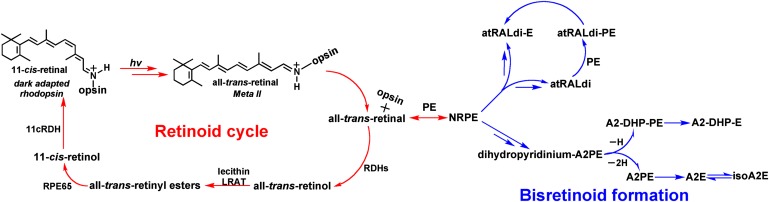
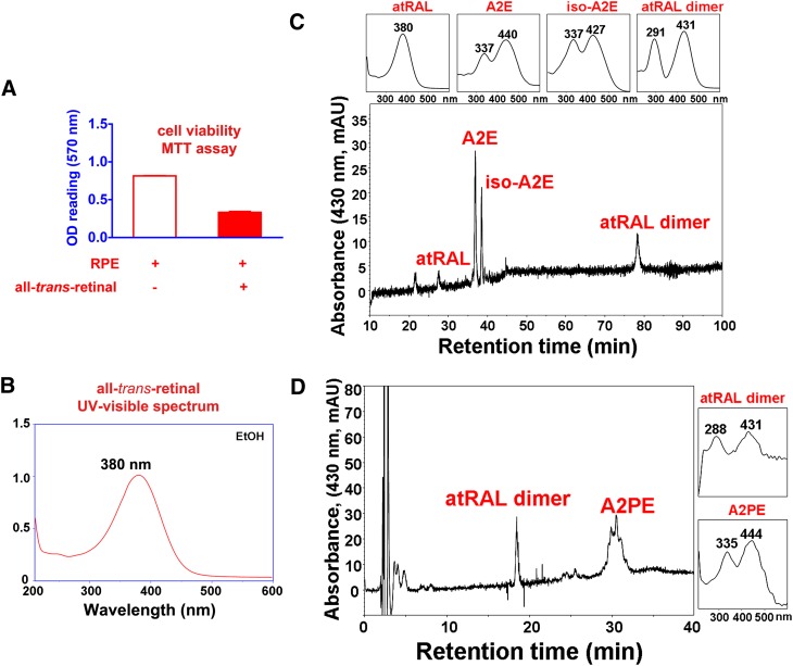
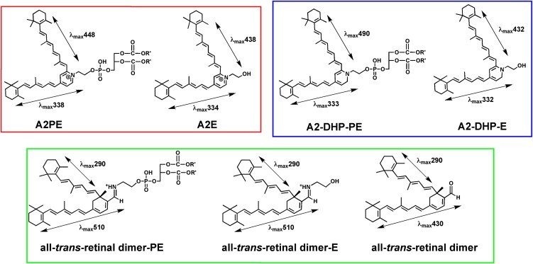
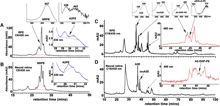
The lipid phase of the photoreceptor outer segment membrane is essential to the photon capturing and signaling functions of rhodopsin. Rearrangement of phospholipids in the bilayer accompanies the formation of the active intermediates of rhodopsin following photon absorption. Furthermore, evidence for the formation of a condensation product between the photolyzed chromophore all-trans-retinal and phosphatidylethanolamine indicates that phospholipid may also participate in the movement of the retinoid in the membrane. The downside of these interactions is the formation of bisretinoid-phosphatidylethanolamine compounds that accumulate in retinal pigment epithelial cells with age and that are particularly abundant in some retinal disorders. The propensity of these compounds to negatively impact on the cells has been linked to the pathogenesis of some retinal disorders including juvenile onset recessive Stargardt disease and age-related macular degeneration.


Figures

Fig. 1.
Fig. 1.
Retinoid cycling in the eye. The visual chromophore 11-cis-retinal forms a covalent Schiff base bond with lysine 296 (Lys 296) of opsin. Vision is initiated when a photon (hv) is captured by 11-cis-retinal; as a result, the chromophore is isomerized to all-trans-retinal. With all-trans-retinal still covalently bound to opsin, the activated pigment transitions to the metarhodopsin II conformation, the Schiff base is hydrolyzed, and all-trans-retinal is reduced to all-trans-retinol by retinol dehydrogenases (RDHs). Alternatively some all-trans-retinal reacts with phosphatidylethanolamine (PE) in the lipid bilayer to form N-retinylidene-PE, which is transported by ABCA4 and then hydrolyzes to release PE and all-trans-retinal. The latter is subsequently reduced to all-trans-retinol. Within the retinal pigment epithelium (RPE) cell, all-trans-retinol is esterified by the enzyme lecithin retinol acyl transferase (LRAT) and is isomerized from the all-trans configuration to the 11-cis-retinol by RPE65. The alcohol is then oxidized by 11-cis retinol dehydrogenase (11cRDH) to 11-cis-retinal. The bisretinoid pathway is initiated when N-retinylidene-PE, rather than hydrolyzing to all-trans-retinal and PE, reacts with a second molecule of all-trans-retinal. A multi-step pathway leads to formation of the intermediate dihydropyridinium-A2PE. Automatic oxidation of dihydropyridinium-A2PE with loss of two hydrogens (−2H) generates A2PE, the immediate precursor of A2E. Loss of one hydrogen (−H) generates A2-dihydropyridine-PE (A2-DHP-PE); phosphate hydrolysis of the latter produces A2-DHP-E. Via an alternative path, all-trans-retinal dimer forms from the condensation of two all-trans-retinal. Reaction all-trans-retinal dimer with PE with formation of a protonated Schiff base linkage generates all-trans-retinal dimer-PE (atRALdi-PE), and phosphate hydrolysis of the latter yields all-trans-retinal dimer-ethanolamine (atRALdi-E).
Fig. 2.
Fig. 2.
All-trans-retinal is generated within photoreceptor cell outer segments upon visual pigment photoisomerization. A: Free all-trans-retinal bears a reactive aldehyde that is toxic to cells. All-trans-retinal (30 microM) was incubated with ARPE-19 cell at 37°C for 16 h after which the cells were incubated with fresh media for 3 days. Cell viability was measured by MTT assay as described in ref. . B: Free all-trans-retinal has an absorbance maxima at 380 nm. C, D: In the presence of lipid bilayers, all-trans-retinal reacts to form several bisretinoid compounds including A2E, isoA2E, all-trans-retinal dimer, and A2PE. All-trans-retinal was incubated with ARPE-19 cells as in A; cells were extracted in chloroform/methanol and analyzed on reverse phase C18 and C8 columns as described in ref. .
Fig. 3.
Fig. 3.
Structures and absorbance maxima (λmax) of the fluorophores that constitute the bisretinoids of retina lipofuscin. Phosphate cleavage of A2PE, A2-DHP-PE, and all-trans-retinal dimer-PE releases A2E, A2-DHP-E, and all-trans-retinal dimer-E, respectively. Absorbance maxima of these bischromophores can be assigned to the shorter and longer side-arms of the molecules. For all-trans-retinal dimer-PE and all-trans-retinal dimer-E, the absorbance generated from the long-arm exhibits a bathochromic shift (red-shift) due to protonation of the imine functional group (— CH = N —).
Fig. 4.
Fig. 4.
Detection of bisretinoids in RPE and neural retina from bovine eyes. Bovine RPE/choroid (A, C) and neural retina (B, D) were extracted with chloroform/methanol and analyzed by reverse-phase HPLC with C8 (A, B) and C18 (C, D) columns and monitoring at 430 and 490 nm. Tissue extracts pooled and concentrated from 15 eyes for RPE analysis and 20 eyes for neural retina analysis. Top insets in A and C: UV-visible absorbance spectra of the indicated bisretinoids. Insets on the right in A–D: Chromatograms expanded between retention times indicated. C: The shorter wavelength absorbance of atRAL dimer-PE presents as ∼265 nm; a strong absorbance in this region from unsaturated fatty acids (such as DHA) in the phosphatidic acid moiety can sometimes mask the 290 nm absorbance generated from the shorter of the two retinoid-derived side-arms of the molecule.
Fig. 5.
Fig. 5.
Fluorescence emission spectra of A2E, all-trans-retinal dimer, and all-trans-retinal dimer-ethanolamine (all-trans-retinal dimer-E), all-trans-retinal dimer-phosphatidylethanolamine (all-trans-retinal dimer-PE). Spectra were recorded at excitation wavelengths of 430 nm (left) and 500 nm (right). Fluorescence intensity is presented in arbitrary units.
Fig. 6.
Fig. 6.
The bisretinoid A2-DHP-PE exhibits fluorescence intensity that is greater than that of A2E when considered relative to the absorbance peaks of each compound. Reverse-phase HPLC chromatogram using C18 column. Overlay of UV-visible absorbance (black) and fluorescence (red; excitation, 430 nm, emission, 600 nm) profiles. AU, absorbance units; a.u., arbitrary units of fluorescence intensity. Insets above, absorbance spectra of A2E, isoA2E, and A2-DHP-PE. B: Chromatogram in A is expanded between retention times 40–50 min; Inset above, absorbance spectra of all-trans-retinal dimer-PE (atRALdi-PE).
Fig. 7.
Fig. 7.
Oxidation of A2E is associated with increased autofluorescence emission. Monitoring of UV-visible absorbance (A) and fluorescence (B) in a mixture of A2E, isoA2E, peroxy-A2E, and peroxy-isoA2E by reverse-phase HPLC. Peroxy-A2/isoA2E was produced by oxidation with endoperoxide of 1,4-dimethylnaphthalene. Presented as absorbance units (A) and arbitrary units of fluorescence intensity (B). Comparison of the absorbance and fluorescence traces for A2E/isoA2E with the same traces for peroxyA2E/peroxy-isoA2E reveals that the fluorescence intensities (peak heights) of bisperoxyA2E and peroxy-isoA2E were considerably increased relative to A2E and isoA2E. C: Structures of A2E, isoA2E, and peroxy-A2E.

References

    1. Hicks D., Sparrow J. R., Barnstable C. J. 1989. Immunoelectron microscopical examination of the surface distribution of opsin in rat rod photoreceptor cells. Exp. Eye Res. 49: 13–29. - PubMed
    1. Fliesler S. J., Anderson R. E. 1983. Chemistry and metabolism of lipids in the vertebrate retina. Prog. Lipid Res. 22: 79–131. - PubMed
    1. McBee J. K., Palczewski K., Baehr W., Pepperberg D. R. 2001. Confronting complexity: the interlink of phototransduction and retinoid metabolism in the vertebrate retina. Prog. Retin. Eye Res. 20: 469–529. - PubMed
    1. Beck M., Siebert F., Sakmar T. P. 1998. Evidence for the specific interaction of a lipid molecule with rhodopsin which is altered in the transition to the active state metarhodopsin II. FEBS Lett. 436: 304–308. - PubMed
    1. Hessel E., Muller P., Hermann A., Hofmann K. P. 2001. Light-induced reorganization of phospholipid in rod disc membrane. J. Biol. Chem. 276: 2538–2543. - PubMed

Publication types

MeSH terms

LinkOut - more resources