. 2009 Aug;21(8):2253-68.
doi: 10.1105/tpc.109.067702. Epub 2009 Aug 7.

Abscisic acid represses growth of the Arabidopsis embryonic axis after germination by enhancing auxin signaling

Affiliations

Abscisic acid represses growth of the Arabidopsis embryonic axis after germination by enhancing auxin signaling

Christophe Belin et al. Plant Cell. 2009 Aug.

Abstract

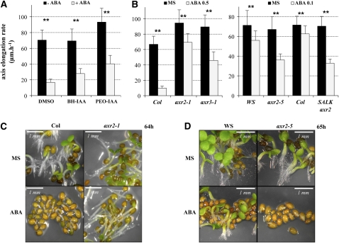
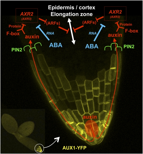
Under unfavorable environmental conditions, the stress phytohormone ABA inhibits the developmental transition from an embryo in a dry seed into a young seedling. We developed a genetic screen to isolate Arabidopsis thaliana mutants whose early seedling development is resistant to ABA. Here, we report the identification of a recessive mutation in AUXIN RESISTANT1 (AUX1), encoding a cellular auxin influx carrier. Although auxin is a major morphogenesis hormone in plants, little is known about ABA-auxin interactions during early seedling growth. We show that aux1 and pin2 mutants are insensitive to ABA-dependent repression of embryonic axis (hypocotyl and radicle) elongation. Genetic and physiological experiments show that this involves auxin transport to the embryonic axis elongation zone, where ABA enhances the activity of an auxin-responsive promoter. We propose that ABA represses embryonic axis elongation by potentiating auxin signaling in its elongation zone. This involves repression of the AUXIN INDUCIBLE (Aux/IAA) gene AXR2/IAA7, encoding a key component of ABA- and auxin-dependent responses during postgerminative growth.


Figures

Figure 1.
Figure 1.
aux1 Mutant Early Seedling Growth Is Resistant to ABA. (A) The pictures show wild-type (Columbia [Col]) and aux1-21, 89 h after seed imbibition in the absence (Murashige and Skoog [MS]) or presence of 0.5 μM ABA (ABA). Bars = 1 mm. (B) The pictures show wild type (Col), aux1-7, and aux1-22, 90 h after seed imbibition in the absence (MS) or presence of 0.5 μM ABA (ABA). Bars = 1 mm. (C) Percentage of emerged green cotyledons for wild type (black bars) and aux1-7 mutant (light-gray bars) was scored 89 h after seed imbibition in the presence of different ABA concentrations, as indicated. Means + sd are shown (n = 2). (D) Time course of the percentage of emerged green cotyledons in wild-type (Col; plain line) and aux1-7 (aux1; dashed line) populations after seed imbibition on medium in the absence (MS; closed symbols) or presence (ABA; open symbols) of 0.2 μM ABA. Means + sd are shown (n = 2).
Figure 2.
Figure 2.
aux1-21 Embryonic Axis Elongation Is Insensitive to ABA. (A) Diagrams and representative pictures showing embryonic axis elongation (Δl) upon endosperm rupture (ER) of the wild type (Col) and aux1-21 (aux1). Seeds were imbibed on normal medium (MS) and transferred at t = 0 h (time of endosperm rupture) to either MS or MS supplemented with 2 μM ABA (ABA). (B) Elongation (Δl) quantification of the experiment described in (A). This graph shows the axis elongation of Col (black bars) and aux1-21 (light-gray bars) between t = 0 h and 3 or 10 h, as indicated, on MS or 2 μM ABA. Standard deviations are shown (n ≥ 20). Asterisks indicate a significant difference between the wild type and the mutant, based on a two-tailed t test (P < 0.01).
Figure 3.
Figure 3.
AUX1 Is Localized in the Plasma Membrane of Lateral Root Cap Cells and Columella Cells during Seed Germination. Confocal microscopy imaging of an AUX1-YFP fusion expressed under the control of the endogenous AUX1 promoter in Col background. (A) Pictures of dissected embryos at 4 stages: 3 h prior to endosperm rupture (ER; i.e., 32 h after seed imbibition), ER (35 h after seed imbibition), and during the elongation of the embryonic axis (37 and 42 h). (B) Magnified pictures of the lateral root cap area of intact germinating seeds at the time of ER (35 h) and during elongation of the embryonic axis (after ER). The arrow indicates testa autofluorescence.
Figure 4.
Figure 4.
Auxin Transport Is Necessary to Mediate ABA-Dependent Repression of Embryonic Axis Elongation. (A) NOA treatment inhibits ABA-dependent repression of embryonic axis elongation. Graph representing the embryonic axis elongation rate of wild type (Col), with (light gray bars) or without 2 μM NOA (dark bars), either in the absence (MS) or presence of 0.5 μM ABA in the medium. (B) Auxin represses embryonic axis elongation. Graph representing the embryonic axis elongation rate of Col (black bars) and aux1-21 (light-gray bars) grown on normal medium (MS) or medium supplemented with synthetic auxins 1 μM NAA or 0.5 μM 2,4-D, as indicated. (C) PIN2 is localized in lateral root cap cells and elongating peripheral (epidermis and cortex) cells during germination. Confocal microscopy imaging of a PIN2-GFP fusion expressed under the control of the endogenous PIN2 promoter in eir1-1 (pin2) background. Whole-embryo pictures at three stages during the axis elongation after endosperm rupture (ER). The fourth picture shows a magnification of the area indicated by the square in the third picture. White arrows indicate testa autofluorescence, and green arrows indicate PIN2-GFP fluorescence. (D) pin2 embryonic axis elongation is insensitive to ABA. Pictures show representative wild-type (Col) and pin2 (eir1-1) germinating seeds, 49 and 64 h after imbibition in presence of 0.5 μM ABA. (E) Embryonic axis elongation rates in Col (black bars) and eir1-1 (light-gray bars) plants grown on normal medium (MS) or medium supplemented with 0.5 μM ABA, 1 μM NAA, or 0.5 μM 2,4-D, as indicated. For (A), (B), and (E), the y axis represents the average embryonic axis elongation rate (μm·h−1). Standard deviations are indicated (n ≥ 15). Asterisks indicate a significant difference between the wild type and the mutant, based on a two-tailed t test (P < 0.01).
Figure 5.
Figure 5.
ABA Potentiates the Effect of Diffusible Auxin NAA to Repress Embryonic Axis Elongation. (A) Average embryonic axis elongation rate of aux1-21 grown on normal medium (-NAA, plain line) or medium supplemented with diffusible synthetic auxin 0.05 μM NAA (NAA 0.05, dashed line), in the presence of different concentrations of ABA. (B) Average embryonic axis elongation rate of aux1-21 grown on normal medium (-ABA, plain line) or medium supplemented with 0.1 μM ABA (+ABA 0.1, dashed line) in the presence of different concentrations of the synthetic auxin NAA. (C) Average embryonic axis elongation rate of wild type (Col, black bars), abi3-9 (white bars), abi4-1 (light-gray bars), and abi5-3 (dark-gray bars) grown on normal medium (MS) or medium supplemented with 0.5 μM ABA, 0.5 μM 2,4-D, or 1 μM NAA, as indicated. Asterisks indicate a significant difference between the wild type and the mutant, based on a two-tailed t test (P < 0.01). For (A) to (C), standard deviations are represented (n ≥ 15).
Figure 6.
Figure 6.
ABA Potentiates Auxin Responses in the Elongation Zone Rather Than Increasing Auxin Levels. (A) Activation of the IAA2 promoter in the elongating zone after endosperm rupture (ER) in response to ABA. Wild-type (Wassilewskija [WS]) lines expressing the uidA gene under the control of the IAA2 promoter were transferred upon ER to normal medium (MS, left panels) or medium supplemented with 2 μM ABA (right panels). Whole seed staining was performed just prior transfer (t = 0 h) and 3 or 10 h thereafter. Red arrows indicate the asymmetric distribution of staining resulting from gravitropic response. (B) ABA-dependent stimulation of transgenic IAA2 promoter requires AUX1. Wild-type (WS) or aux1 (aux1-100) seeds expressing the uidA gene under the control of the IAA2 promoter were sown on medium with 0.5 μM ABA. Whole-seed staining was performed 41 and 110 h after imbibition for each genotype. (C) Global auxin levels in early seedlings in response to ABA. Auxin levels were measured in the same samples used in (A) and in dry seeds (DS). (D) Auxin levels in Col and abi5-3 early seedlings, transferred upon endosperm rupture (ER) to normal medium (MS) or medium supplemented with 2 μM ABA. Auxin levels were measured as in (C) just prior to transfer (t0) and 3 or 10 h thereafter. Auxin levels in dry seeds (DS) are also shown. In (C) and (D), means + sd are shown (n = 3).
Figure 7.
Figure 7.
Aux/IAA Turnover Is Involved in ABA-Dependent Repression of Embryonic Axis Elongation. (A) Average embryonic axis elongation rate of wild type (Col) in absence (DMSO) or presence of auxin antagonists (BH-IAA and PEO-IAA, 50 μM each) competing with endogenous IAA to bind TIR1/AFB F-box proteins. Seeds were imbibed on normal medium (MS) and transferred to different media, as indicated, either in the absence (- ABA, black bars) or presence of 2 μM ABA (+ ABA, light-gray bars). (B) Quantification of embryonic axis elongation in axr2/iaa7 and axr3/iaa17 mutants. Left: average axis elongation rate of wild type (Col), axr2-1, and axr3-1, as indicated, in absence (MS, black bars) or presence of 0.5 μM ABA (light-gray bars). Right: average axis elongation rate of wild type (Col and WS), axr2-5 (in WS ecotype), and SALK axr2/iaa7 line (in Col ecotype), as indicated, in the absence (MS, black bars) or presence of 0.1 μM ABA (light-gray bars). (A) And (B) Standard deviations are indicated (n ≥ 13). Asterisks indicate a significant difference between untreated (-ABA or MS) and ABA-treated (+ABA or ABA) conditions, based on a two-tailed t test (P < 0.01). (C) Pictures of seeds from wild type (Col) and axr2-1 64 h after imbibition in the absence (MS) or presence of 0.5 μM ABA. (D) Pictures of seeds from wild type (WS) and axr2-5 65 h after imbibition in the absence (MS) or presence of 0.5 μM ABA.
Figure 8.
Figure 8.
AXR2/IAA7 and AXR3/IAA17 Upstream Sequences Are Transcriptionally Active in the Peripheral Elongation Zone; ABA Represses AXR2/IAA7 and AXR3/IAA17 mRNA Expression. (A) Localization of AXR2/IAA7 promoter activity during early seedling growth. Pictures of wild-type transgenic lines expressing the uidA gene under the control of the AXR2/IAA7 promoter. Whole-seed staining is visible only 42 to 45 h after seed imbibition on normal germination medium. Squares represent the areas magnified in the right panels. Arrows indicate the hypocotyl/root junction (collet). (B) Localization of AXR3/IAA17 promoter activity during early seedling growth. Pictures of wild-type transgenic line expressing the uidA gene under the control of the AXR3/IAA17 promoter. Whole-seed staining was performed 37 h after seed imbibition on normal germination medium. Squares represent the areas magnified in the left panels. (C) RNA gel blot analysis of AXR2/IAA7 and AXR3/IAA17 mRNA levels in wild-type (Col) and aux1-21 (aux1) during embryonic axis elongation. Seeds were transferred upon endosperm rupture (t0; i.e., 34 h after seed imbibition) to normal medium (MS) or medium supplemented with 2 μM ABA for 1.5, 3, or 10 h, as indicated. rRNA is shown as a loading control.
Figure 9.
Figure 9.
A Model of ABA and Auxin Crosstalk for the Repression of Embryonic Axis Elongation. This model is drawn on a magnified confocal micrograph of the embryonic axis tip of a wild-type line expressing ProAUX1:AUX1-YFP (Figure 4; see Methods). Auxin (red) is distributed from the radicle tip to the elongation zone via the influx facilitator AUX1 located in the root cap (yellow) and the efflux carrier PIN2 polarly localized in the elongating epidermal and cortical cells (green). In the elongation zone, auxin activates its signaling pathway (red) by binding to TIR1/AFB F-box proteins, promoting the degradation of the Aux/IAA repressors AXR2/IAA7 and possibly AXR3/IAA17. This would, in turn, activate unknown transcriptional regulator ARFs to repress cell elongation. ARFs appear in brackets because their involvement in ABA- and auxin-dependent repression of embryonic axis elongation remains speculative. We propose that ABA (blue) potentiates the auxin pathway by strongly decreasing the mRNA levels of the Aux/IAA repressor AXR2/IAA7 (and possibly AXR3/IAA17).

Similar articles

Cited by

References

    1. Abel, S., Nguyen, M.D., and Theologis, A. (1995). The PS-IAA4/5-like family of early auxin-inducible mRNAs in Arabidopsis thaliana. J. Mol. Biol. 251: 533–549. - PubMed
    1. Alonso, J.M., et al. (2003). Genome-wide insertional mutagenesis of Arabidopsis thaliana. Science 301: 653–657. - PubMed
    1. Beaudoin, N., Serizet, C., Gosti, F., and Giraudat, J. (2000). Interactions between abscisic acid and ethylene signaling cascades. Plant Cell 12: 1103–1115. - PMC - PubMed
    1. Benjamins, R., and Scheres, B. (2008). Auxin: The looping star in plant development. Annu. Rev. Plant Biol. 59: 443–465. - PubMed
    1. Bennett, M.J., Marchant, A., Green, H.G., May, S.T., Ward, S.P., Millner, P.A., Walker, A.R., Schulz, B., and Feldmann, K.A. (1996). Arabidopsis AUX1 gene: A permease-like regulator of root gravitropism. Science 273: 948–950. - PubMed

Publication types

MeSH terms

LinkOut - more resources