. 2009 Sep;136(17):2965-75.
doi: 10.1242/dev.036616.

MARCKS modulates radial progenitor placement, proliferation and organization in the developing cerebral cortex

Affiliations

MARCKS modulates radial progenitor placement, proliferation and organization in the developing cerebral cortex

Jill M Weimer et al. Development. 2009 Sep.

Abstract

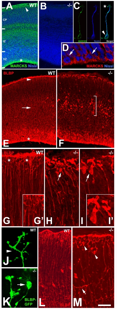
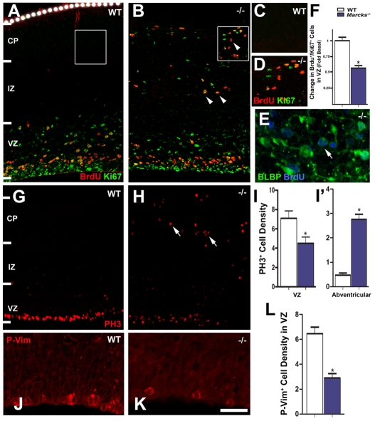
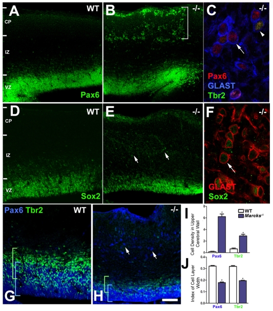
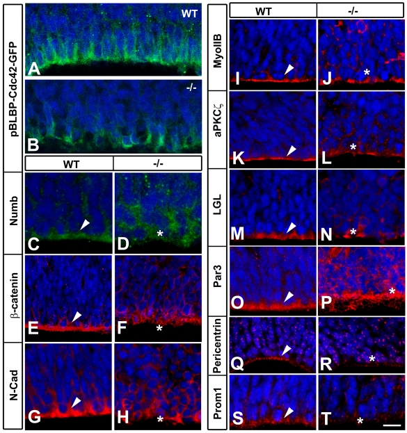
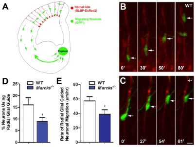
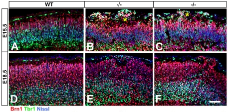
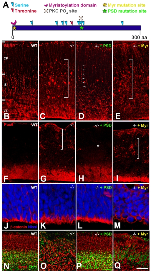
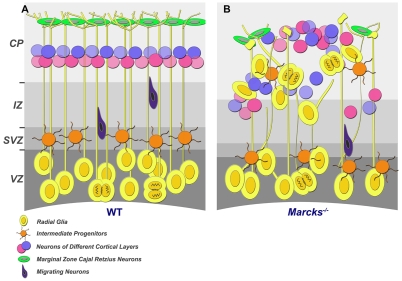
The radial glial cells serve as neural progenitors and as a migratory guide for newborn neurons in the developing cerebral cortex. These functions require appropriate organization and proliferation of the polarized radial glial scaffold. Here, we demonstrate in mice that the myristoylated alanine-rich C-kinase substrate protein (MARCKS), a prominent cellular substrate for PKC, modulates radial glial placement and expansion. Loss of MARCKS results in ectopic collection of mitotically active radial progenitors away from the ventricular zone (VZ) in the upper cerebral wall. Apical restriction of key polarity complexes [CDC42, beta-catenin (CTNNB1), N-cadherin (CDH2), myosin IIB (MYOIIB), aPKCzeta, LGL, PAR3, pericentrin, PROM1] is lost. Furthermore, the radial glial scaffold in Marcks null cortex is compromised, with discontinuous, non-radial processes apparent throughout the cerebral wall and deformed, bulbous, unbranched end-feet at the basal ends. Further, the density of radial processes within the cerebral cortex is reduced. These deficits in radial glial development culminate in aberrant positioning of neurons and disrupted cortical lamination. Genetic rescue experiments demonstrate, surprisingly, that phosphorylation of MARCKS by PKC is not essential for the role of MARCKS in radial glial cell development. By contrast, the myristoylation domain of MARCKS needed for membrane association is essential for MARCKS function in radial glia. The membrane-associated targeting of MARCKS and the resultant polarized distribution of signaling complexes essential for apicobasal polarity may constitute a critical event in the appropriate placement, proliferation and organization of polarized radial glial scaffold in the developing cerebral cortex.


Figures

Fig. 1.
Fig. 1.
Disruption in radial glial scaffold in Marcks-/- cerebral cortex. (A-D) MARCKS is widely expressed in the E15.5 cerebral cortex (A, green). Prominent MARCKS expression is apparent in the apical and basal ends of the cerebral wall, where the radial progenitor cell soma (asterisk, A) and end-feet (arrowhead, A) are located. MARCKS immunoreactivity is absent in the Marcks-/- brain (B). An isolated radial glial cell co-immunostained with anti-MARCKS and radial-glia-specific nestin (blue) antibodies indicates prominent MARCKS expression in the cell soma (arrowhead, C) and at the tips of the radial processes (asterisk, C). High magnification view of the VZ shows apical expression of MARCKS in actively dividing radial progenitors (arrows, D). Nuclei are counterstained with Nissl (blue) in A,B,D. (E-K) Anti-BLBP labeling outlines the normal radial glial scaffold in the wild-type (WT) cortex: radial glial cell soma in the VZ (asterisk, E), radial processes spanning the cerebral wall (arrow, E) and end-feet under the pial surface (arrowhead, E). In Marcks-/- cortex, an ectopic band of BLBP+ cells is present in the IZ/CP of the cerebral wall, away from the VZ (bracket, F). Compared with the branched end-feet in control cortices (asterisks, G), radial glial end-feet are distorted and club-like in Marcks-/- cortex (arrows; H,I). Insets (G′,I′) show end-feet from WT (G′) and Marcks-/- (I′) cortex at high magnification. Labeling of radial glial end-feet using in utero electroporation of BLBP promoter-GFP plasmids into WT (J) and Marcks-/- (K) cortices further illustrates this alteration [i.e. branched WT end-feet (arrowhead, J) versus club-like end-feet in Marcks-/- radial glia (arrow, K)]. (L,M) As corticogenesis progresses (E18.5), the BLBP+ radial processes in WT cortices continue to expand across the extent of the cerebral wall (L), whereas MARCKS-deficient radial glia frequently display misoriented radial processes (arrow, M) and bulbous end-feet at or near the pial surface (arrowheads, M). Scale bar in M: 100 μm for A,B; 30 μm for D,L,M; 80 μm for E,F; 20 μm for G-I; 10 μm for J,K. CP, cortical plate; IZ, intermediate zone; VZ, ventricular zone.
Fig. 2.
Fig. 2.
MARCKS deletion leads to ectopic placement and proliferation of radial progenitors. (A,B) BrdU (red) and Ki67 (green) labeling of WT cortex (E15.5) indicates actively proliferating radial progenitors are primarily within the VZ/SVZ (A). By contrast, ectopic, mitotically active BrdU+/Ki67+ cells are found within the IZ/CP in Marcks-/- cortex (arrowheads, B). (C,D) High magnification images of boxed regions of the CP in A and B, respectively, illustrate the ectopic presence of proliferating progenitors in Marcks-/- cortex. (E) These ectopically proliferating cells are positive for BLBP (green), a marker for radial progenitors (arrow). BrdU labeling is blue. (F) BrdU and Ki67 co-labeling indicates that the fraction of actively cycling progenitors (BrdU+/Ki67+) in the VZ is reduced in Marcks-/- cortex (blue bar). (G-L) Immunolabeling with PH3 (G,H) or anti-phospho-vimentin (P-Vim; J,K) antibodies reveals a uniform line of mitotically active, radial progenitors confined to the ventricular surface in WT cortex (G,J). In Marcks-/- brains, fewer PH3+ and P-Vim+ cells are localized to the ventricular surface (H,I,K,L). M-phase, PH3+ progenitors are seen displaced throughout the IZ/CP in Marcks-/- cortex (arrows, H). Quantification of the increase in ectopically placed [abventricular (IZ/CP)] PH3+ progenitors (I). Data shown are mean cell density (104/mm2) ±s.e.m.; asterisks indicate significance when compared with controls at P≤0.01. Scale bar in K: 50 μm for A,B,G,H; 40 μm for C,D,J,K; 30 μm for F. The dotted line in A indicates the pial surface. CP, cortical plate; IZ, intermediate zone; VZ, ventricular zone.
Fig. 3.
Fig. 3.
Molecular identity of ectopic progenitors in Marcks-/- cortex. (A-F) Immunolabeling of E15.5 radial progenitors in WT (A,D) and Marcks-/- (B,E) cortex with anti-PAX6 and anti-SOX2 antibodies illustrates the presence of ectopic PAX6+ (bracket, B) and SOX2+ (arrows, E) progenitors within the IZ/CP in Marcks-/- cortex. These ectopic PAX6+, TBR2+ and SOX2+ progenitors within the IZ/CP co-label with radial-glia-specific anti-GLAST antibodies (arrows, C,F; arrowhead in panel C indicates a GLAST+ cell labeled with both PAX6 and TBR2). (G,H) Radial and intermediate progenitors in WT and Marcks-/- cortex were immunolabeled with anti-PAX6 and anti-TBR2 antibodies. Compared with WT (G), PAX6+ and TBR2+ cells are displaced away from the VZ/SVZ region in Marcks-/- cortex (arrows, H). (I,J) Quantification of PAX6+ and TBR2+ cell density (104/mm2) in the upper cerebral wall indicates the extent of ectopic placement of progenitors in Marcks-/- cortex (I). The width of PAX6+ and TBR2+ cell layers in the VZ is significantly reduced in Marcks-/- cortex (compare brackets in G and H; quantified in J). Data shown are mean±s.e.m.; asterisk indicates significance when compared with controls at P≤0.01. Scale bar in H: 100 μm for A,B,D,E; 20 μm for C,F; 50 μm for G,H; 75 μm for I. CP, cortical plate; IZ, intermediate zone; VZ, ventricular zone.
Fig. 4.
Fig. 4.
Changes in the expression of apical polarity markers in Marcks-/- radial progenitors. (A,B) CDC42-GFP expression in WT (A) and Marcks-/- (B) radial progenitors shows that normal apical localization of CDC42-GFP is disrupted in Marcks null radial progenitors (B). (C-T) Immunolabeling of E15.5 cortices with anti-NUMB (C,D), β-catenin (E,F), anti-N-cadherin (N-cad; G,H), anti-myosin IIB (MyoIIB; I,J), anti-aPKCζ (K,L), anti-LGL (M,N), anti-PAR3 (O,P), anti-pericentrin (Q,R) and anti-prominin1 (PROM1; S,T) antibodies demonstrates a loss of polarized localization of these proteins away from the apical surface of the Marcks-/- radial progenitors (compare areas indicated by arrowheads and asterisks). Punctate labeling with anti-pericentrin (Q-R) indicates isolated centrosomes. Nuclei are counterstained with Nissl (blue). Scale bar in T: 10 μm for A-D; 20 μm for E-T.
Fig. 5.
Fig. 5.
Marcks deletion disrupts radial glia-guided migration. WT and Marcks-/- E15.5 cortices were electroporated with BLBP promoter-dsRed2 plasmids to label radial glia (red). (A)GFP+ explants from the medial ganglionic eminence of Dlx5/6-CIE mice were then overlaid onto electroporated slices to enable neuronal migration on radial glial processes. Time-lapse imaging of these assays was used to monitor radial glia-neuron interactions. (B) Illustration of a migrating neuron (arrow, green) along a WT radial glial process (red). Time elapsed between observations is indicated in minutes. (C) Illustration of the altered migration of a neuron (arrow, green) on Marcks-/- radial processes (red). (D) Quantification of radial glia-neuron interactions in these assays indicates that fewer neurons initiate contact with and utilize MARCKS-deficient radial glial processes as migratory guides. (E) Compared with WT controls, the average rate of radial-glial-guided migration is significantly lower on Marcks-/- radial glia. Data shown are mean±s.e.m.; asterisk indicates significance when compared with controls at P≤0.01. Scale bar in C: 7.5 μm for B,C.
Fig. 6.
Fig. 6.
Defective neuronal lamination in MARCKS-deficient cortex. (A) Immunolabeling with anti-BRN1 (green) and anti-TBR1 (red) antibodies demarcates distinct layers in E15.5 WT CP. (B,C) BRN1+ and TBR1+ neurons are intermixed and laminar organization is disrupted in Marcks-/- cortex (asterisks). (D-F)This neuronal layering defect persists at later developmental stages (E18.5; compare D with E and F). Scale bar in F: 50 μm for A-F.
Fig. 7.
Fig. 7.
Myristoylation domain of MARCKS is crucial for its function. (A) A schematic of full-length MARCKS. Transgenic lines expressing MARCKS with a mutated PSD or myristoylation moiety (Myr) were crossed onto a Marcks-/- background to determine the functionally crucial domains of MARCKS. Regions mutated in the transgenic lines are indicated (asterisks). (B,C) Immunolabeling with an anti-BLBP antibody indicates a normal radial glial scaffold in WT cortex (B) and a disrupted radial glial scaffold with ectopic BLBP+ radial progenitors in Marcks-/- cortex (bracket, C). Anti-PAX6 labeling also indicates ectopic placement of radial progenitors in Marcks-/- cortex (F,G; bracket). The PSD-domain mutant MARCKS is able to rescue the ectopic BLBP+ (D) and PAX6+ (H) radial progenitor distribution in the upper IZ/CP. Arrows in D point to radial processes in Marcks-/- + PSD cortex as seen in WT controls. Similarly, the asterisk in H indicates a lack of ectopic PAX6+ cells in Marcks-/- + PSD cortex. Moreover, PSD-domain mutant MARCKS also rescues the disrupted apical localization of β-catenin (J-L), and the cortical lamination defects (N-P). The Myr-domain mutant MARCKS, however, fails to rescue the ectopic placement of BLBP+ (bracket, E) or PAX6+ (bracket, I) progenitors, the loss of apical-restricted expression of β-catenin in the VZ (M), or cortical lamination defects (Q). Scale bar in Q: 80 μm for B-E; 40 μm for F-I; 100 μm for K-N; 20 μm for O-R; 70 μm for S-V. CP, cortical plate; IZ, intermediate zone; VZ, ventricular zone.
Fig. 8.
Fig. 8.
Role of MARCKS in radial glial organization and cortical development. (A) MARCKS facilitates the proper placement and organization of the radial glial scaffold. This is essential for the laminar organization of neurons in the neocortex. (B) Loss of MARCKS leads to ectopic placement of radial progenitors within the IZ/CP, a disorganized radial glial scaffold with abnormal glial end-feet and process orientation, and aberrant cortical lamination. MARCKS may serve as an anchor for the polarized spatial localization of crucial signaling complexes within radial glial progenitors, and thus promotes proper radial glial development and cortical organization. CP, cortical plate; IZ, intermediate zone; SVZ, subventricular zone; VZ, ventricular zone.

Similar articles

Cited by

References

    1. Aderem, A. (1992a). The MARCKS brothers: a family of protein kinase C substrates. Cell 71, 713-716. - PubMed
    1. Aderem, A. (1992b). Signal transduction and the actin cytoskeleton: the roles of MARCKS and profilin. Trends Biochem. Sci. 17, 438-443. - PubMed
    1. Anton, E. S., Cameron, R. S. and Rakic, P. (1996). Role of neuron-glial junctional domain proteins in the maintenance and termination of neuronal migration across the embryonic cerebral wall. J. Neurosci. 16, 2283-2293. - PMC - PubMed
    1. Arbuzova, A., Schmitz, A. A. and Vergeres, G. (2002). Cross-talk unfolded: MARCKS proteins. Biochem. J. 362, 1-12. - PMC - PubMed
    1. Ayala, R., Shu, T. and Tsai, L. H. (2007). Trekking across the brain: the journey of neuronal migration. Cell 128, 29-43. - PubMed

Publication types

MeSH terms

Substances