. 2009 Oct;77(10):4621-30.
doi: 10.1128/IAI.00415-09. Epub 2009 Aug 10.

Cytotoxicity and secretion of gamma interferon are carried out by distinct CD8 T cells during Mycobacterium tuberculosis infection

Affiliations

Cytotoxicity and secretion of gamma interferon are carried out by distinct CD8 T cells during Mycobacterium tuberculosis infection

Thorbjorg Einarsdottir et al. Infect Immun. 2009 Oct.

Abstract

The host immune response is generally sufficient to contain Mycobacterium tuberculosis infection. It does not, however, efficiently prevent subsequent infection with M. tuberculosis or provide sterilizing immunity. While the understanding of the immune response generated against this pathogen is incomplete, improvements have been achieved due to advances in immunological tools. In this study, we analyzed the multifunctional nature of primary and memory CD8 T-cell responses generated during murine M. tuberculosis infection. We generated a recombinant M. tuberculosis strain expressing ovalbumin (OVA) epitopes in order to expand the peptides for the detection of CD8 T cells during M. tuberculosis infection and enable us to use OVA-specific reagents. Our results indicate that the majority of M. tuberculosis-specific CD8 T cells are limited to either cytotoxicity or the secretion of gamma interferon (IFN-gamma), with cytotoxicity being far more prevalent than IFN-gamma secretion. Memory CD8 T cells responded earlier and reached higher levels in the lungs than naïve CD8 T cells, as was expected. They were, however, less cytotoxic and secreted less IFN-gamma than newly primed CD8 T cells, suggesting that one factor contributing to bacterial persistence and lack of sterilizing immunity may be the low quality of memory cells that are generated.


Figures

FIG. 1.
FIG. 1.
Generation and screening of M. tuberculosis strain ova. (A) Map of pMH94CFP10ova integration vector, drawn with Vector NTI suite 8 (InforMax, Inc.). CFP10 and the upstream sequence were inserted into pMH94, followed by the OVA sequence. The plasmid lacks an origin of replication in M. tuberculosis and is lost unless it is integrated in a site-specific manner into the genome by the integrase via the attP site. Recombinant colonies were selected based on kanamycin resistance (KanR). (B) PCR screening for integration of pMH94CFP10ova into M. tuberculosis. Lane 1 shows a 100-bp DNA ladder (Invitrogen). Lanes 2 (WT M. tuberculosis) and 4 (M. tuberculosis strain ova) show positive controls, screening for endogenous copies of CFP10 (225 bp). Lanes 3 (WT M. tuberculosis) and 5 (M. tuberculosis strain ova) screen for pMH94CFP10ova, with primers specific for the ends of the CFP10-OVA fusion gene (312 bp; indicated by the arrow). The figure has been edited to remove irrelevant lanes. (C) ELISPOT assay measuring IFN-γ secretion in response to M. tuberculosis (Mtb), endogenous T-cell epitopes (GAP and ESAT-6), OVA CD8 T-cell epitopes, and background levels (media). The data are the means ± standard errors of means from testing results for four mice. OVA-specific CD8 T-cell responses induced by M. tuberculosis strain ova (Mtb-ova) were statistically different (**, P < 0.01) than responses induced by WT M. tuberculosis, while other responses were not statistically different.
FIG. 2.
FIG. 2.
M. tuberculosis strain ova induces similar GAP- and OVA-specific responses. Based on the percent tetramer-positive (Tet+) CD8 T cells (see Fig. S1A and B in the supplemental material) and live cell numbers (data not shown), the numbers of tetramer-positive cells in the lungs (A) and lymph nodes (B) were calculated. The frequencies of tetramer-specific cells during primary (1′) and secondary (2′) infections were significantly different at 0, 2, 3, and 4 weeks postinfection for OVA-specific cells (o*) and at 0, 2, and 4 weeks postinfection for GAP-specific cells (g*). The frequencies of OVA- and GAP-specific cells were significantly different at 8 weeks (lungs) and 28 weeks (lymph nodes) after primary infection (go1*) and 4 weeks (lungs) after secondary infection (go2*). (C) Bacterial load in the lungs. Primary and secondary infections were statistically different at 3 and 4 weeks postinfection (P < 0.01). Data are the means ± standard errors of the means from test results for 4 (A and B) to 12 (C) mice per time point.
FIG. 3.
FIG. 3.
IFN-γ secretion and degranulation are carried out by distinct populations of cells. Based on GAP- and OVA-specific CD8 T-cell numbers shown in Fig. 2A and percent IFN-γ+ (IFNg+) and CD107+ cells (see Fig. S3A and B in the supplemental material), numbers of cells per organ were calculated for primary (A and B) and secondary (C and D) infections. (A and C) GAP; (B and D) OVA. The data are the means ± standard errors of the means from test results for four mice per time point. There was a significant difference in the frequency (P < 0.05) and number (P < 0.05) of IFN-γ+ CD107+ cells between primary and secondary infections for GAP-specific cells at 2 weeks postinfection and OVA-specific CD8 T cells at 2 and 4 weeks postinfection.
FIG. 4.
FIG. 4.
Functional assays for GAP- and OVA-specific cytotoxicity and IFN-γ secretion. (A) ELISPOT assay, where lung cells were incubated with DCs pulsed with GAP or OVA peptide (SIINFEKL) for 48 h. There was no background level of IFN-γ (IFNg) secretion, measured by incubating cells with either T-cell media or unpulsed, uninfected DCs (data not shown). Data are means ± standard errors of the means from test results for four mice per time point. There was no statistical significance between primary (1′) and secondary (2′) infections, except for OVA-specific cells at 8 weeks postinfection (P < 0.05). (B) In vivo cytotoxicity assay in the lung. Data are the averages from test results using four mice per group. (C) GAP-specific cytotoxicity versus the frequency of CD107+ GAP-specific CD8 T cells in the lung. There was a significant correlation (P < 0.05) between cytotoxicity and CD107 surface expression during primary infection, as measured by the Spearman nonparametric test, while there was not a significant correlation during secondary infection.
FIG. 5.
FIG. 5.
Blocking PD-1 signal in vivo does not give IFN-γ+ CD107+ cells. A total of 5 × 10³ OT-I cells (CD45.2) were adoptively transferred to CD45.1 mice, which were subsequently infected with M. tuberculosis strain ova. Using anti-CD45.2 antibodies, responses of these cells were examined over the course of infection. (A) Percent CD45.2+ OT-I+ cells expressing PD-1; (B) geometric MFI of PD-1 staining in panel A. (C and D) Mice treated with anti-PD-1 and anti-PD-L1 antibodies, or with PBS, for 4 weeks. Ab, antibody; a-, anti-. (C) GAP+ CD8 T cells expressing IFN-γ (IFNg) and surface CD107. (D) Bacterial load in the lung and lymph nodes. Data are the means ± standard errors of the means from test results for three or four mice per experimental group.
FIG. 6.
FIG. 6.
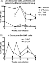
CD8 T cells express perforin, while granzyme B expression can be induced. (A) Cells were stained, without incubation, for perforin (fluorescein isothiocyanate, clone dG9) and granzyme B (GrB), using fixative and a protocol from Apo-Active 3 (Cell Technology, Mountain View, CA). 1′, primary infection; 2′, secondary infection. (B) Cells were stained for granzyme B after a 5-h incubation, as described in Materials and Methods. Data are the means ± standard errors of the means from test results for three or four mice per experimental group.
FIG. 7.
FIG. 7.
IFN-γ+ CD107+ cells secrete more IFN-γ and degranulate more than cells limited to either function. The geometric MFIs of CD107+ and IFN-γ+ GAP-specific cells were analyzed. IFN-γ (IFNg) MFI (A) or CD107 MFI (B) of cells that both secrete IFN-γ and degranulate or are limited to either function. Data are the means ± standard errors of the means from test results for four mice per time point. There were significant differences in both IFN-γ secretion and degranulation between primary (1′) and secondary (2′) infections (P < 0.05) at 3 and 4 weeks postinfection. IFN-γ+ CD107+ cells expressed significantly more IFN-γ at 4 weeks postinfection than IFN-γ+ CD107 cells (P < 0.01), while there was no significant difference in degranulation between IFN-γ+ CD107+ and IFN-γ CD107+ cells.

References

    1. Agnellini, P., P. Wolint, M. Rehr, J. Cahenzli, U. Karrer, and A. Oxenius. 2007. Impaired NFAT nuclear translocation results in split exhaustion of virus-specific CD8+ T cell functions during chronic viral infection. Proc. Natl. Acad. Sci. USA 104:4565-4570. - PMC - PubMed
    1. Algood, H. M. S., P. L. Lin, D. Yankura, A. Jones, J. Chan, and J. L. Flynn. 2004. TNF influences chemokine expression of macrophages in vitro and that of CD11b+ cells in vivo during Mycobacterium tuberculosis infection. J. Immunol. 172:6846-6857. - PubMed
    1. Barber, D. L., E. J. Wherry, D. Masopust, B. Zhu, J. P. Allison, A. H. Sharpe, G. J. Freeman, and R. Ahmed. 2006. Restoring function in exhausted CD8 T cells during chronic viral infection. Nature 439:682-687. - PubMed
    1. Betts, M. R., J. M. Brenchley, D. A. Price, S. C. De Rosa, D. C. Douek, M. Roederer, and R. A. Koup. 2003. Sensitive and viable identification of antigen-specific CD8+ T cells by a flow cytometric assay for degranulation. J. Immunol. Methods 281:65-78. - PubMed
    1. Bibb, L. A., and G. F. Hatfull. 2002. Integration and excision of the Mycobacterium tuberculosis prophage-like element, phiRv1. Mol. Microbiol. 45:1515-1526. - PubMed

Publication types