. 2009 Aug 31;206(9):1913-27.
doi: 10.1084/jem.20082297. Epub 2009 Aug 10.

Cancer cell-derived microparticles bearing P-selectin glycoprotein ligand 1 accelerate thrombus formation in vivo

Affiliations

Cancer cell-derived microparticles bearing P-selectin glycoprotein ligand 1 accelerate thrombus formation in vivo

Grace M Thomas et al. J Exp Med. .

Abstract

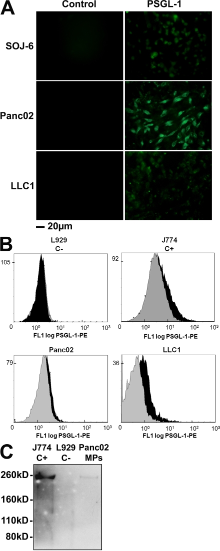
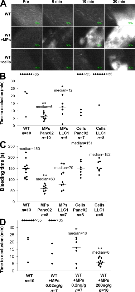
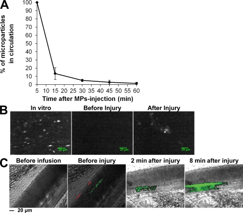
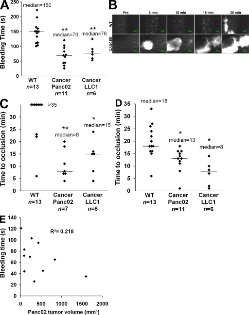
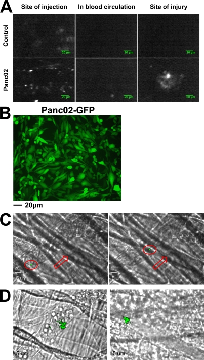
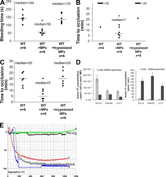
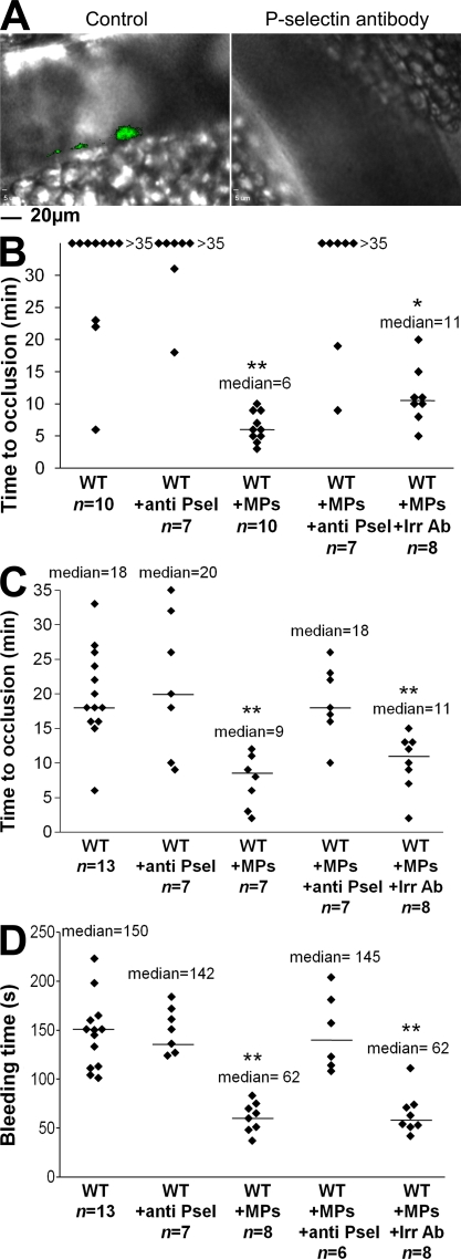
Recent publications have demonstrated the presence of tissue factor (TF)-bearing microparticles (MPs) in the blood of patients suffering from cancer. However, whether these MPs are involved in thrombosis remains unknown. We show that pancreatic and lung cancer cells produce MPs that express active TF and P-selectin glycoprotein ligand 1 (PSGL-1). Cancer cell-derived MPs aggregate platelets via a TF-dependent pathway. In vivo, cancer cell-derived MPs, but not their parent cells, infused into a living mouse accumulate at the site of injury and reduce tail bleeding time and the time to occlusion of venules and arterioles. This thrombotic state is also observed in mice developing tumors. In such mice, the amount of circulating platelet-, endothelial cell-, and cancer cell-derived MPs is increased. Endogenous cancer cell-derived MPs shed from the growing tumor are able to accumulate at the site of injury. Infusion of a blocking P-selectin antibody abolishes the thrombotic state observed after injection of MPs or in mice developing a tumor. Collectively, our results indicate that cancer cell-derived MPs bearing PSGL-1 and TF play a key role in thrombus formation in vivo. Targeting these MPs could be of clinical interest in the prevention of thrombosis and to limit formation of metastasis in cancer patients.


Figures

Figure 1.
Figure 1.
Pancreatic and lung cancer cell lines shed MPs that express TF in vitro. (A) Flow cytometry analysis showing annexin V–FITC labeling of SOJ-6, Panc02, and LLC1 cell–derived MPs. Negative controls were realized without addition of calcium. (B) Immunofluorescence microscopy of cancer cells using an anti-TF antibody. (C) Western blot analysis of protein extracts from SOJ-6–derived MPs with a specific anti-TF antibody. All of the experiments were independently performed three times.
Figure 2.
Figure 2.
Pancreatic and lung cancer cell–derived MPs express PSGL-1. (A) Immunofluorescence microscopy of SOJ-6, Panc02, and LLC1 cells with an antibody directed against PSGL-1 and an Alexa Fluor 488–conjugated secondary antibody. Incubation with the secondary antibody alone served as a negative control. (B) Flow cytometric analysis of J774 (C+), L929 (C−), Panc02-, and LLC1-derived MPs labeled both with annexin V–FITC and with an anti–PSGL-1 PE-conjugated antibody. PSGL-1+ MPs are represented by the black histogram. Negative controls were obtained by excluding calcium from the annexin V labeling and in the presence of an irrelevant PE-conjugated IgG (gray histogram). (C) Western blot analysis of Panc02-derived MPs (Panc02 MPs) and J774 (C+) and L929 (C−) cells with a specific anti–PSGL-1 antibody. All of the experiments were independently performed three times.
Figure 3.
Figure 3.
Cancer cell–derived MPs accelerate thrombus formation in vivo. Thrombus formation was assessed after infusion of Alexa Fluor 660–conjugated anti-CD41 antibody into wild-type mice in the presence of Panc02-derived MPs (MPs Panc02, 0.2 µg of MP-associated proteins/g/mouse), LLC1-derived MPs (MPs LLC1, 0.08 µg of MP-associated proteins/g/mouse), or Panc02 cells (Cells Panc02, 5,000 cells/g/mouse) or LLC1 cells (Cells LLC1, 5,000 cells/g/mouse). Injury of the mesenteric vessels was induced with 10% FeCl3 for 5 min. (A) Representative composite images of fluorescence depicting thrombus formation of labeled platelet accumulation (white) on mesenteric vessels (Pre, before injury). (B) Time to vessel occlusion reported in minutes for mesenteric arterioles in wild-type mice (n = 10 thrombi in 10 mice), wild-type mice infused with Panc02-derived MPs (n = 10 thrombi in 7 mice), or with LLC1-derived MPs (n = 6 thrombi in 6 mice), Panc02 cells (n = 7 thrombi in 7 mice), or LLC1 cells (n = 8 thrombi in 8 mice). (C). Bleeding time in seconds determined in wild-type mice (n = 13 mice), wild-type mice infused with Panc02-derived MPs (n = 8 mice), or with LLC1-derived MPs (n = 7 mice), Panc02 cells (n = 8 mice), or LLC1 cells (n = 8 mice). (D) Times to arteriole occlusion were observed in wild-type mice (n = 10 thrombi in 10 mice) and in wild-type mice infused with different concentrations of Panc02-derived MPs (MPs 0.02 ng/g/mouse, n = 7 thrombi in 6 mice; MPs 0.2 ng/g mouse, n = 7 thrombi in 7 mice; and MPs 200 ng/g mouse, n = 10 thrombi in 9 mice). Horizontal bars indicate median values. Experiments were independently performed 10 times (A and B) and at least six times (C and D). **, P < 0.01; *, P < 0.05.
Figure 4.
Figure 4.
Cancer cell–derived MPs accumulate at the site of injury in mice. (A) Half-life of DiO-labeled Panc02-derived MPs in the blood circulation of a healthy mouse (n = 4 mice, performed independently) reported as the mean ± SD. (B) Imaging of DiD-labeled Panc02-derived MPs using fibered fluorescence microscopy. Panc02-derived MPs were labeled with DiD (left) before infusion into the blood circulation of a living mouse. Although no fluorescent signal was detected in the microcirculation before injury (middle), MPs were detected accumulating at the site of FeCl3-induced injury (right). Images are representative of six injuries performed in three mice, independently. (C) Intravital videomicroscopy visualization of mesenteric vessels. DiO-labeled MPs (depicted in green; 0.2 µg of MP-associated proteins/g/mouse) and their DiD-labeled parental cells (depicted in red; 5,000 cells/g/mouse) were simultaneously infused into a recipient mouse. Circulating MPs and cells were detected in the microcirculation before injury (second from left). Note that no fluorescent signal was detected before infusion of MPs and cells (left). The experiment was independently performed three times in three mice.
Figure 5.
Figure 5.
Characterization of a thrombotic state in mice developing tumors. Kinetics of thrombosis in wild-type mice or in mice developing Panc02-derived (Cancer Panc02) or LLC1-derived (Cancer LLC1) tumors. (A) Bleeding times were determined in wild-type mice (n = 13 mice), mice developing a Panc02 tumor (n = 11 mice), or mice developing an LLC1 tumor (n = 6 mice). (B) Thrombus formation was studied after infusion of Alexa Fluor 660–conjugated anti-CD41 Fab fragment and Syto 62 in wild-type mice and in mice developing tumors. Injury was induced in mesenteric vessels by topical application of 10% FeCl3 for 5 min. Representative fluorescence images depicting the kinetics of thrombus formation of labeled platelet accumulation (white) on mesenteric arterioles (Pre, before injury). (C and D) Time to vessel occlusion reported in minutes for mesenteric arterioles (n = 13, 7, and 6 thrombi for WT, Cancer Panc02, and Cancer LLC1, respectively; C) and venules (n = 13, 11, and 6 thrombi for WT, Cancer Panc02, and Cancer LLC1, respectively; D). One injury was performed per mouse. (E) Bleeding times (in seconds) in function of the tumor volume (in mm3) in Panc02 tumor–bearing mice (n = 11). The linear coefficient of determination value is represented by R2. Horizontal bars in A, C, and D indicate median values. Experiments were independently performed at least six times (A–D) and 11 times (E). **, P < 0.01; *, P < 0.05.
Figure 6.
Figure 6.
Endogenous cancer cell–derived MPs accumulate at the site of thrombus formation in vivo. (A) Qdot-labeled Panc02 cells (2 × 10⁶ cells) or Qdots alone (Control) were injected subcutaneously into the right flank of a mouse. 1 wk later, fluorescent signals were detected at the site of injection (left), and in the mesentery before (middle panel) and after (right) FeCl3-induced injury. (B) Fluorescence microscopy of Panc02 cells overexpressing GFP before subcutaneous injection into wild-type mice. (C and D) 2 × 10⁵ Panc02 cells overexpressing GFP were injected subcutaneously into the right flank of a mouse. 5 wk later, the cremaster was isolated and fluorescent circulating GFP-labeled MPs were detected in the blood microcirculation (red arrow, direction of the blood flow; red circle, GFP microparticle; C) and accumulating at the site of injury in venules (D, left) and arterioles (D, right). All images are representative of three independent experiments observed for nine thrombi formed in three mice.
Figure 7.
Figure 7.
Endogenous cancer-derived MPs accelerate thrombus growth through a TF-dependent pathway. (A–C) Kinetics of thrombosis in mice perfused with control Panc02 MPs or trypsinized Panc02 MPs (18,000 MPs/g/mouse) compared with wild-type mice. (A) Bleeding time of wild-type mice and of mice perfused with control MPs (WT+MPs) or trypsinized MPs (WT+trypsinized MPs; n = 6 mice for each group). Time to vessel occlusion reported in minutes for mesenteric arterioles (B) and venules (C; n = 6 thrombi in six mice for each group). Injury of the mesenteric vessels was induced by topical application of 10% FeCl3 for 5 min. Horizontal bars indicate median values. **, P < 0.01; *, P < 0.05. (D) Measurement of TF activity on the surface of cancer cells and MPs in pmol/cell−1 and pmol/MP−1 (left) or in pmol/mm2 (right), reported as the mean ± SD. (E) Aggregation of 2.5 × 10⁸ human washed platelets/ml induced by addition of thrombin (Curve 1, red), SOJ-6 MPs (1 µg of MP-associated proteins/ml) in the absence (Curve 2, black) or presence of 5% vol/vol (final) of PPP (Curve 3, blue), factor VII–depleted PPP (Curve 4, green), or fibrinogen-deficient PPP (Curve 5, gray). Experiments were independently performed six times (A–C) and three times (D and E).
Figure 8.
Figure 8.
Endogenous cancer-derived MPs accelerate thrombus formation in a P-selectin–dependent manner. (A) 2 × 10⁵ Panc02 cells overexpressing GFP were injected subcutaneously into the right flank of a mouse. 5 wk later, chemical injury of the mesentery was induced, and fluorescence was detected in the absence (left, Control) or presence of circulating P-selectin blocking antibody (right, P-selectin antibody; n = 6 thrombi in three mice). (B–D). Kinetics of thrombosis in wild-type mice, wild-type mice infused with Panc02 MPs (WT+MPs, 0.2 µg of MP-associated proteins/g/mouse), or with Panc02 MPs plus 2 µg/g/mouse of anti P-selectin blocking antibody (WT+MPs+anti P-sel). Controls were performed by infusion of anti–P-selectin antibody into wild-type mice (WT+anti P-sel) or by infusing an irrelevant antibody into wild-type mice perfused with MPs (WT+MPs+Irr Ab). Injury of the mesenteric vessels was chemically induced, and the time to vessel occlusion for arterioles (n = 10 for WT, n = 7 for WT+anti Psel, n = 10 for WT+MPs, n = 7 for WT+MPs+anti Psel, and n = 8 for WT+MPs+Irr Ab; B) and veinules (n = 13 for WT, n = 7 for WT+anti Psel, n = 7 for WT+MPs, n = 7 for WT+MPs+anti Psel, and n = 8 for WT+MPs+Irr Ab; C) was measured. (D) Bleeding times were determined for all conditions studied (n = 13 for WT, n = 7 for WT+anti Psel, n = 8 for WT+MPs, n = 6 for WT+MPs+Anti Psel, and n = 8 for WT+MPs+Irr Ab). One thrombus was performed per mouse. Horizontal bars designate median values. Experiments were independently performed three times (A) and at least seven times (B–D). **, P < 0.01; *, P < 0.05.

Similar articles

Cited by

References

    1. Abid Hussein M.N., Meesters E.W., Osmanovic N., Romijn F.P., Nieuwland R., Sturk A. 2003. Antigenic characterization of endothelial cell-derived microparticles and their detection ex vivo.J. Thromb. Haemost. 1:2434–2443 - PubMed
    1. Berckmans R.J., Neiuwland R., Böing A.N., Romijn F.P., Hack C.E., Sturk A. 2001. Cell-derived microparticles circulate in healthy humans and support low grade thrombin generation.Thromb. Haemost. 85:639–646 - PubMed
    1. Bertram J.S., Janik P. 1980. Establishment of a cloned line of Lewis Lung Carcinoma cells adapted to cell culture.Cancer Lett. 11:63–73 - PubMed
    1. Blom J.W., Osanto S., Rosendaal F.R. 2006a. High risk of venous thrombosis in patients with pancreatic cancer: a cohort study of 202 patients.Eur. J. Cancer. 42:410–414 - PubMed
    1. Blom J.W., Vanderschoot J.P., Oostindiër M.J., Osanto S., van der Meer F.J., Rosendaal F.R. 2006b. Incidence of venous thrombosis in a large cohort of 66,329 cancer patients: results of a record linkage study.J. Thromb. Haemost. 4:529–535 - PubMed

Publication types