. 2009 Aug 31;206(9):1929-40.
doi: 10.1084/jem.20090896. Epub 2009 Aug 10.

Decreased TNF-alpha synthesis by macrophages restricts cutaneous immunosurveillance by memory CD4+ T cells during aging

Affiliations

Decreased TNF-alpha synthesis by macrophages restricts cutaneous immunosurveillance by memory CD4+ T cells during aging

Elaine Agius et al. J Exp Med. .

Abstract

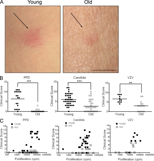
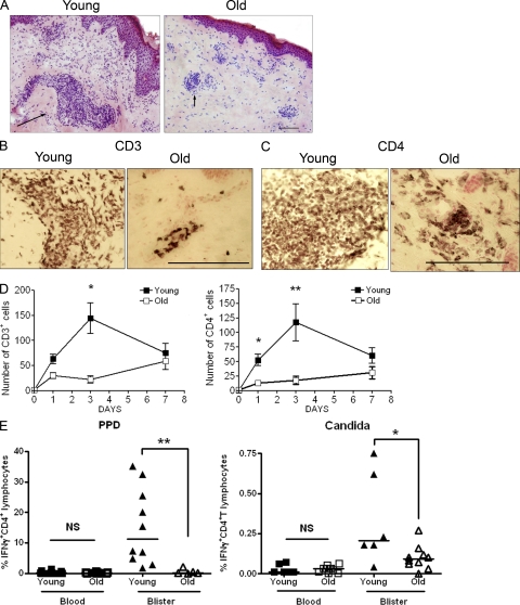
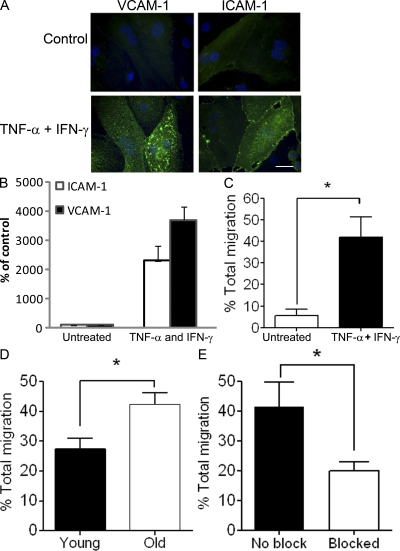
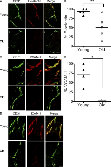
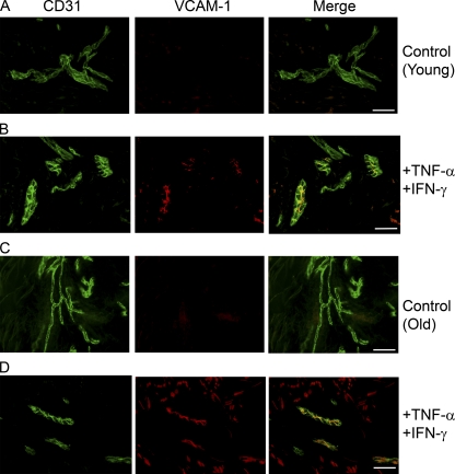
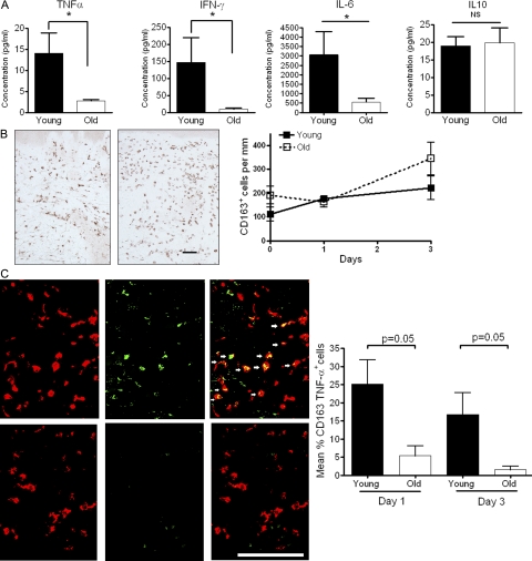
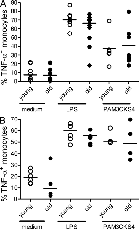
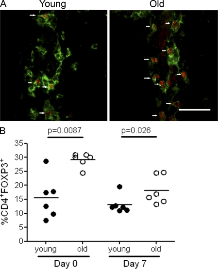
Immunity declines during aging, however the mechanisms involved in this decline are not known. In this study, we show that cutaneous delayed type hypersensitivity (DTH) responses to recall antigens are significantly decreased in older individuals. However, this is not related to CC chemokine receptor 4, cutaneous lymphocyte-associated antigen, or CD11a expression by CD4(+) T cells or their physical capacity for migration. Instead, there is defective activation of dermal blood vessels in older subject that results from decreased TNF-alpha secretion by macrophages. This prevents memory T cell entry into the skin after antigen challenge. However, isolated cutaneous macrophages from these subjects can be induced to secrete TNF-alpha after stimulation with Toll-like receptor (TLR) 1/2 or TLR 4 ligands in vitro, indicating that the defect is reversible. The decreased conditioning of tissue microenvironments by macrophage-derived cytokines may therefore lead to defective immunosurveillance by memory T cells. This may be a predisposing factor for the development of malignancy and infection in the skin during aging.


Figures

Figure 1.
Figure 1.
Cutaneous DTH responses are reduced in old individuals. Young and old volunteers were injected intradermally with 0.1 ml of 10 U/ml tuberculin PPD (n = 20 young, 32 old), 0.02 ml Candin skin antigen test (n = 40 young, 34 old), or 0.02 ml VZV skin test antigen (n = 11 young, 15 old). (A) A photograph of the injection site 3 d after PPD injection. Bar, 1 cm. (B) Induration diameter, erythema index, and palpability of the lesion were assessed and graded at day 3, and a clinical score was assigned based on these parameters (***, P < 0.0001; **, P < 0.001; Mann-Whitney test). (C) Freshly isolated PBMCs were stimulated with PPD (n = 18 young, 28 old), C. albicans (n = 23 young, 19 old), or VZV (n = 7 young, 10 old) in vitro for 5–6 d. Proliferation was assessed by [3H]thymidine incorporation and plotted against corresponding clinical score. Each symbol represents the result from one individual.
Figure 2.
Figure 2.
Cellular infiltrate at the site of cutaneous DTH response is reduced in the old. Young and old volunteers were injected with 0.02 ml Candin skin antigen test and 5-mm punch biopsies were performed on days 1, 3, or 7 after injection. (A) Hematoxylin and eosin staining of skin sections from one young and one old individual on day 3 after Candin injection showing perivascular infiltrates (black arrow). The results are representative stains of three young and three old subjects that were tested. Day 3 biopsies were stained for CD3 (B) and CD4 (C) using an indirect immunoperoxidase immunohistochemical technique. The results shown are representative of five young and four old subjects and the quantification of this data are included in D. (D) The number of CD3+ and CD4+ T cells within perivascular infiltrates was determined by counting the number of positive cells within the five largest perivascular infiltrates per skin section. Mean ± SEM of 4–7 individuals per time point is shown (*, P = 0.01; **, P = 0.007; Mann-Whitney test; n = 20 old, 22 young with 4–7 volunteers per time point). (E) The percentage IFN-γ–producing antigen specific CD4+ T lymphocytes in old blisters is significantly reduced compared with young (n = 10 young, 5 old; **, P = 0.001, for PPD specific cells and n = 6 young, 10 old; *, P = 0.04 for C. albicans). Each symbol represents the result of a single subject. Horizontal bars represent the median. Bars, 100 µm.
Figure 3.
Figure 3.
CD4+ T cells from old volunteers show adequate ability to migrate across the dermal endothelial monolayer in vitro. (A) Photomicrographs immunostaining of HDMEC monolayer for VCAM-1 and ICAM-1 (green) on unactivated (top) and activated (bottom) endothelium. Endothelial monolayers were activated with TNF-α (110,000 U/ml) and IFN-γ (100 U/ml) for 24 h before addition of T cells. Bar,10 µm. (B) Graph showing ICAM-1 and VCAM-1 up-regulation upon HDMEC activation. Results were expressed as mean ± SEM of triplicate wells. (C) Bar graph showing increased total CD4+ T cell migration across stimulated HDMEC compared with unstimulated endothelium. To evaluate the level of migration, CD4+ T cells isolated from young and old volunteers were plated onto confluent dermal endothelial monolayers for 4 h at 37°C and 5% CO2 (n = 3 young, 3 old). (D) The percentage total migration for old versus young CD4+ T cells across stimulated endothelium was significantly different (Mann Whitney *, P = 0.02; n = 4 young, 4 old). (E) Graph showing effect of adding blocking antibodies to ICAM-1, VCAM-1 and E-selectin on total CD4+ lymphocyte migration (Paired t test *P = 0.04). Results were expressed as mean ± SEM of three separate experiments.
Figure 4.
Figure 4.
Dermal endothelium in old individuals shows reduced expression of adhesion molecules. Histological sections (6 µm) from skin injected with C. albicans (n = 5 young, 6 old) at day 3 after injection were stained with antibody to CD31 to highlight dermal capillary loops and endothelial adhesion molecules E-selectin VCAM-1 and ICAM-1. (A) Double immunofluorescence staining of representative biopsies for one young and one old individual showing CD31 (green) and E-selectin (red). Capillary loops expressing adhesion molecules appear yellow (merge). This staining was repeated on biopsies of skin 3 d after C. albicans injection in five different young and five different old subjects. The collective results are shown in B. The graph shows percentage of capillary loops expressing E-selectin and the horizontal line indicates the mean (**, P = 0.008, young vs. old, Mann-Whitney test). (C) VCAM-1 (red) and CD31 (green) expression by skin biopsies taken 3 d after C. albicans injection. Representative staining of one young and one old subject is shown. These experiments were performed on four young and five old subjects, and the collective results are shown in D. Graph shows the percentage of capillary loops expressing VCAM-1; each symbol represents an individual, and lines indicate the mean (*, P = 0.015, young vs. old). (E) ICAM-1 (red) and CD31 (green) immunofluorescence staining of skin biopsies from one representative young and one representative old individual 3 d after C. albicans injection. These experiments are representative results of staining that was performed in five young and five old subjects. Bars, 100 µm.
Figure 5.
Figure 5.
Old endothelium is not defective and can up-regulate adhesion molecules in vitro. Young and old normal skin explants were cultured in RPMI + 10% human serum or in RPMI + 10% human serum supplemented with TNF-α (110,000 U/ml) and IFN-γ (100 U/ml) for 16 h at 37°C and 5% CO2. Histological sections from skin explants were stained with antibodies to CD31 and E-selectin, ICAM-1, and VCAM-1. The results shown represent one young (A and B) and one old (C and D) subject showing VCAM-1 (red) and CD31 (green) staining. These experiments were performed in a total of three different young and three different old subjects that showed similar results. Bars, 100 µm.
Figure 6.
Figure 6.
Levels of TNF-α are reduced in blister fluid and in CD163+ macrophages in old skin. (A) Skin suction blisters were raised over the site of C. albicans antigen injection at day 3. Levels of TNF-α, IFN-γ, IL-10 and IL-6 were measured in the blister fluid using cytometric bead arrays. The results shown are the mean and SEM of 6 young and 10 old subjects for each cytokine tested. A significant reduction in the level of TNF-α, IFN-γ, and IL-6 was noted in the old group compared with the young group (Mann-Whitney test; *, P = 0.01 for TNF-α and IFN-γ; *, P = 0.03 for IL-6). The number of CD163+ cells within the dermis was determined by indirect immunoperoxidase staining of 6-µm tissue sections. (B, left) Representative staining of one young and one old subject 3 d after C. albicans injection. We investigated a total of five young and six old volunteers, and the collective results are shown (B, right). We also investigated the presence of CD163+ macrophages at different times after C. albicans injection (B, right). Graph shows mean number of CD163+ macrophages before injection (n = 5 young, 5 old), day 1 (n = 5 young, 7 old), and day 3 after injection (n = 5 young, 6 old). The data represent the mean ± SEM. Bar, 100 µm. Double immunofluorescence staining of TNF–α (green) and CD163 (red) in skin biopsies 3 d after C. albicans injection. Results shown are representative of one young and one old subject (C, left). These experiments were performed in three young and three old subjects and the collective results showing the mean and SEM of the percentage of CD163+ macrophages producing TNF-α per perivascular infiltrate are shown in (C, right). Bar, 100 µm.
Figure 7.
Figure 7.
Macrophages isolated from the skin of old donors secrete TNF-α in response to TLR signals in vitro. Cells removed from skin suction blisters 48 h after C. albicans injection were stimulated with LPS (100 ng/ml; Sigma-Aldrich), Pam3CSK4 (1 µg/ml), or medium as a control for 4 h in the presence of GolgiStop, and then stained for CD14, CD163, and TNF–α. PBMCs were stimulated in parallel. (A) Graph shows percentage of TNF-α–secreting CD14+ cells after in vitro stimulation of PBMCs with medium alone or LPS (n = 11 young, 11 old) and Pam3CSK4 (n = 6 young, 7 old). Each symbol represents the results from one individual. The percentage of CD14+ cells producing TNF-α after in vitro stimulation of blister cells with LPS (n = 6 young, 7 old) and Pam3CSK4 (n = 3 young, 4 for old). (B) Each symbol represents the results from one individual.
Figure 8.
Figure 8.
Numbers of CD4+Foxp3+ T reg cells are increased in the skin of old subjects. (A) Double immunofluorescence staining of representative biopsies from young and old normal skin; CD4+ (green) and Foxp3+ (red) cells in a perivascular lymphocytic infiltrate. CD4+Foxp3+ cells are indicated by a white arrow. Bar, 100 µm. (B) Percentage of CD4+ cells expressing Foxp3 per perivascular infiltrate counted. The five largest perivascular infiltrates present in the upper and mid-dermis were selected for analysis for each subject. Each symbol represents an average of five perivascular infiltrates counted for each individual (n = 5–7 subjects per time point, line indicates the mean).

References

    1. Adamson P., Etienne S., Couraud P.O., Calder V., Greenwood J. 1999. Lymphocyte migration through brain endothelial cell monolayers involves signaling through endothelial ICAM-1 via a rho-dependent pathway.J. Immunol. 162:2964–2973 - PubMed
    1. Alon R., Kassner P.D., Carr M.W., Finger E.B., Hemler M.E., Springer T.A. 1995. The integrin VLA-4 supports tethering and rolling in flow on VCAM-1.J. Cell Biol. 128:1243–1253 - PMC - PubMed
    1. Balato A., Balato N., Di Costanzo L., Ayala F. 2008. Contact sensitization of older patients in an academic department in Naples, Italy.Dermatitis. 19:209–212 - PubMed
    1. Berg E.L., Yoshino T., Rott L.S., Robinson M.K., Warnock R.A., Kishimoto T.K., Picker L.J., Butcher E.C. 1991. The cutaneous lymphocyte antigen is a skin lymphocyte homing receptor for the vascular lectin endothelial cell-leukocyte adhesion molecule 1.J. Exp. Med. 174:1461–1466 - PMC - PubMed
    1. Berlin C., Bargatze R.F., Campbell J.J., von Andrian U.H., Szabo M.C., Hasslen S.R., Nelson R.D., Berg E.L., Erlandsen S.L., Butcher E.C. 1995. alpha 4 integrins mediate lymphocyte attachment and rolling under physiologic flow.Cell. 80:413–422 - PubMed

Publication types

Substances