. 2009 Nov;29(21):5963-73.
doi: 10.1128/MCB.01268-08. Epub 2009 Aug 10.

Modulation of Rho guanine exchange factor Lfc activity by protein kinase A-mediated phosphorylation

Affiliations

Modulation of Rho guanine exchange factor Lfc activity by protein kinase A-mediated phosphorylation

David Meiri et al. Mol Cell Biol. 2009 Nov.

Abstract

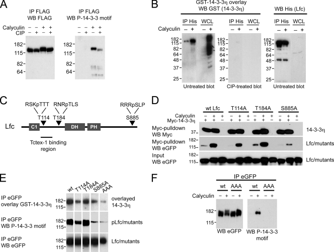
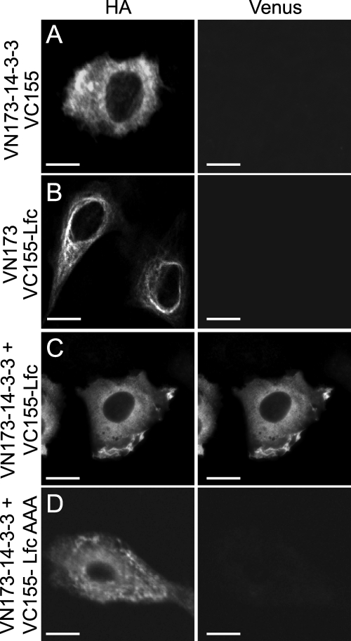
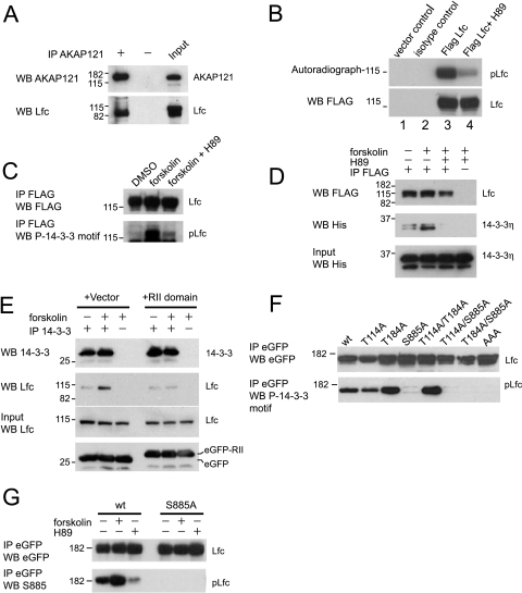
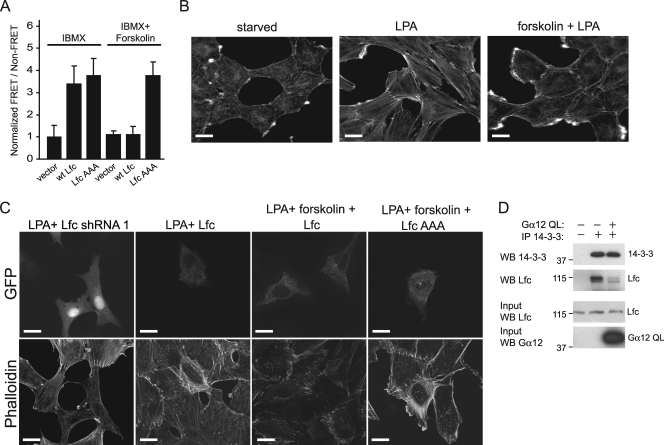
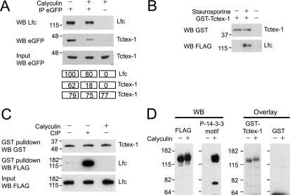
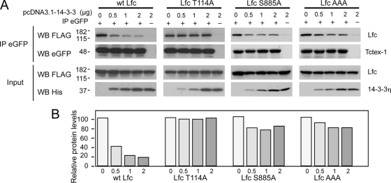
Lfc is a guanine nucleotide exchange factor (GEF) for Rho that demonstrates an unusual ability to associate with microtubules. While several phosphorylated residues have been detected in the Lfc polypeptide, the mechanism(s) by which phosphorylation regulates the exchange activity of Lfc remains unclear. We confirm that Lfc is a phosphorylated protein and demonstrate that 14-3-3 interacts directly and in a phosphorylation-dependent manner with Lfc. We identify AKAP121 as an Lfc-binding protein and show that Lfc is phosphorylated in an AKAP-dependent manner by protein kinase A (PKA). Forskolin treatment induced 14-3-3 binding to Lfc and suppressed the exchange activity of wild-type Lfc on RhoA. Importantly, a mutant of Lfc that is unable to associate with 14-3-3 proteins was resistant to inhibition by forskolin. Tctex-1, a dynein motor light chain, binds to Lfc in a competitive manner with 14-3-3.


Figures

FIG. 1.
FIG. 1.
Lfc is a phosphoprotein and associates directly with 14-3-3η. (A) 293T cells overexpressing FLAG-tagged Lfc were treated with calyculin or dimethylsulfoxide (DMSO) vehicle for 30 min immediately prior to lysis. Anti-FLAG immunoprecipitates (IP) were left untreated or were treated with CIP and immunoblotted for FLAG-Lfc or phosphorylated 14-3-3 binding motifs. WB, Western blotting. (B) 14-3-3η binds to Lfc directly, and the binding is phosphorylation dependent. 293T cells transfected with a His-tagged Lfc construct or untransfected cells were treated with calyculin (or vehicle) for 30 min, and immunoprecipitated His-Lfc protein or whole-cell lysate (WCL) was electrophoresed and transferred to PVDF. Membranes were incubated with or without CIP overnight prior to the overlay of recombinant GST-14-3-3η or immunoblotted for His-Lfc. (C) Domain structure of Lfc showing three predicted 14-3-3-binding sites and their amino acid sequences. The bar represents the Tctex-1-binding region (residues 87 to 151). (D) Myc-14-3-3η coimmunoprecipitation performed using lysates from 293T cells expressing eGFP-tagged Lfc, Lfc T114A, Lfc T184A, or Lfc S885A treated with calyculin or left untreated. Myc-14-3-3η complexes were electrophoresed, transferred to PVDF, and immunoblotted with anti-Myc or anti-eGFP to detect 14-3-3η and Lfc, respectively. wt, wild type. (E) Far-Western analysis of immunoprecipitated, eGFP-tagged Lfc, Lfc T114A, Lfc T184A, Lfc S885A, or Lfc-AAA prepared from calyculin-treated 293T cells. Membranes were probed with purified recombinant GST-14-3-3η (upper) or immunoblotted for phosphorylated 14-3-3 binding motifs (middle) or total eGFP-tagged Lfc protein levels (bottom). (F) Lfc is phosphorylated at sites in addition to 14-3-3 consensus sites. 293T cells transfected with eGFP-tagged Lfc or Lfc-AAA (AAA) were left untreated or were treated with calyculin. Immunoprecipitated Lfc and Lfc-AAA complexes were electrophoresed and immunoblotted for total levels of Lfc protein (anti-eGFP) and phosphorylated 14-3-3 binding motifs (anti-P-14-3-3 motif).
FIG. 2.
FIG. 2.
Lfc interacts with 14-3-3η in cells. 14-3-3η and Lfc were cloned into pCMV-HA vectors proximal to N-terminal (VN173) or C-terminal (VC155) fragments of Venus, respectively, and were introduced into Rat2 cells. Transfected cells were imaged by confocal microscopy. (A) Coexpression of VN173-14-3-3 and VC155 (vector without insert). (B) Coexpression of VN173 (vector without insert) and VC155-Lfc. (C) Coexpression of VN173-14-3-3 and VC155-Lfc. (D) Coexpression of VN173-14-3-3 and VC155-Lfc-AAA. Bars represent 10 μM.
FIG. 3.
FIG. 3.
Lfc associates with AKAP121 and is phosphorylated by PKA. (A) Lfc interacts with AKAP121. Endogenous AKAP121 was immunoprecipitated (IP) from NIH 3T3 cells, and immunoprecipitates were immunoblotted for AKAP121 and Lfc. WB, Western blotting. (B) Lfc immune complex kinase assay. pFLAG-Lfc was immunoprecipitated from transfected 293T cells, washed, and incubated with [γ-32P]ATP. The products of the kinase assay were visualized by autoradiography. Transfected cells were treated with vehicle or the PKA inhibitor H89 prior to lysis. (C) PKA phosphorylates Lfc. 293T cells were transfected with FLAG-tagged Lfc, and cells were treated with DMSO or 30 μM H89 for 1 h prior to the addition of 10 μM forskolin for 30 min. Anti-FLAG immunoprecipitates were immunoblotted with anti-FLAG antibodies (upper) or anti-phospho-14-3-3 motifs antibodies (lower). (D) Forskolin treatment enhances 14-3-3 binding to Lfc. 293T cells were transfected with FLAG-tagged Lfc and His-tagged 14-3-3η and pretreated with DMSO vehicle or 30 μM H89 for 1 h prior to the addition of 10 μM forskolin to culture media. FLAG-tagged complexes were immunoprecipitated and subjected to Western blotting for FLAG-Lfc and the associated His-14-3-3η. (E) Forskolin-induced 14-3-3 binding to Lfc is AKAP dependent. NIH 3T3 cells were transfected with eGFP or the eGFP-tagged RII-binding domain of AKAP121 and were left untreated or treated with 10 μM forskolin for 30 min. Endogenous 14-3-3 complexes were immunoprecipitated and immunoblotted for 14-3-3 or Lfc. (F) PKA phosphorylates Lfc on residues T114 and S885. Anti-eGFP immunoprecipitates prepared from 293T cells expressing eGFP-tagged wild-type (wt) Lfc or Lfc mutants and treated with 10 μM forskolin for 30 min were immunoblotted for eGFP or anti-phospho-14-3-3 motif antibodies. (G) PKA phosphorylates Lfc at S885. 293T cells were transfected with wild-type eGFP-tagged Lfc, Lfc S885A, or Lfc T114/S885A and treated with 10 μM forskolin for 30 min or 30 μM H89 for 1 h. eGFP-tagged complexes were immunoprecipitated and subjected to Western blotting with anti-eGFP or anti-S885 Lfc antibody.
FIG. 4.
FIG. 4.
PKA inhibits RhoA activation by Lfc and stress fiber assembly. (A) RhoA-Raichu assay of Lfc exchange activity. NIH 3T3 cells transfected with the Raichu probe and His-tagged wild-type (wt) Lfc or Lfc-AAA were treated with IBMX alone or IBMX and forskolin for 10 min, and lysates were prepared to measure normalized fluorescence at 528 and 480 nm. Experiments were performed three times, and results are expressed as a ratio of basal FRET ± standard errors. (B) Forskolin inhibits stress fiber assembly. Rat2 cells were grown on slides and starved of serum for 24 h. Cells were fixed or pretreated with or without 10 μM forskolin for 30 min prior to 1 μM LPA treatment for 30 min. Coverslips were stained with phalloidin to visualize actin filaments. (C) LPA signaling to the actin cytoskeleton requires active Lfc. Rat2 cells were transfected with short hairpin RNA, eGFP Lfc, or eGFP-Lfc-AAA and starved for 24 h. Cells were fixed or pretreated with 10 μM forskolin or left untreated for 30 min prior to 1 μM LPA treatment for 30 min. Coverslips were stained with phalloidin to visualize actin filaments. Bars represent 10 μM. (D) Gα12 modulates 14-3-3 binding to Lfc. Gα12 inhibits the interaction of Lfc with 14-3-3 proteins. 293T cells were transfected with pcDNA3.1 vector or pcDNA3.1-Gα12 Q231L (Gα12 QL). Endogenous 14-3-3 complexes were immunoprecipitated (IP) from lysates and separated by SDS-PAGE. After transfer to PVDF, membranes were subjected to Western blotting (WB) for 14-3-3 and Lfc.
FIG. 5.
FIG. 5.
Dephosphorylation of Lfc promotes Tctex-1 binding. (A) Endogenous Lfc complexes immunoprecipitated (IP) from Rat2 cells stably expressing eGFP-Tctex-1 left untreated or treated with calyculin A were immunoblotted for Lfc (top) or associated eGFP-Tctex-1 (middle). The intensity of the bands was determined by densitometry using ImageJ Prime software and are presented under the bands. WB, Western blotting. (B) Phosphorylation regulates Tctex-1 binding to Lfc. 293T cells expressing FLAG-tagged Lfc were treated with 0.5 μM staurosporine for 1 h, and lysates were incubated with immobilized GST-Tctex-1. GST complexes were immunoblotted for FLAG-Lfc and GST-Tctex-1. (C) Lfc dephosphorylation promotes Tctex-1 binding. Lysates prepared from FLAG-Lfc-transfected 293T cells were incubated with or without CIP, or cells were treated with calyculin prior to lysis. Lysates were incubated with GST-Tctex-1, and immobilized GST complexes were immunoblotted for GST-Tctex-1 (upper) and FLAG-Lfc (middle). (D) Tctex-1 overlay assay of nonphosphorylated and phosphorylated Lfc. Cells transfected with FLAG-Lfc were treated with calyculin for 15 min, and FLAG-Lfc immunocomplexes were electrophoresed and immunoblotted for total Lfc protein levels (first gel) or phosphorylated 14-3-3 binding motifs (second gel), or membranes were overlaid with purified recombinant GST-Tctex-1 (third gel) or GST alone (fourth gel). GST-Tctex-1 associated directly with FLAG-Lfc equally independent of phosphorylation status.
FIG. 6.
FIG. 6.
14-3-3 and Tctex-1 bind to Lfc in a mutually exclusive manner. (A) 293T cells were transfected with FLAG-tagged Lfc, Lfc T114A, Lfc S885A, Lfc-AAA, and eGFP-tagged Tctex-1 in the presence of increasing amounts of His-tagged 14-3-3η. eGFP immunocomplexes and input lysates were immunoblotted for FLAG-Lfc, eGFP-Tctex-1, and His-14-3-3η. IP, immunoprecipitation; WB, Western blotting; wt, wild type. (B) The intensity of the bands of variant FLAG-tagged Lfc from panel A were determined (by densitometry using ImageJ Prime software) by dividing the observed signal of each band with that of input FLAG-tagged Lfc.

Similar articles

Cited by

References

    1. Aghazadeh, B., W. E. Lowry, X. Y. Huang, and M. K. Rosen. 2000. Structural basis for relief of autoinhibition of the Dbl homology domain of proto-oncogene Vav by tyrosine phosphorylation. Cell 102:625-633. - PubMed
    1. Aijaz, S., F. D'Atri, S. Citi, M. S. Balda, and K. Matter. 2005. Binding of GEF-H1 to the tight junction-associated adaptor cingulin results in inhibition of Rho signaling and G1/S phase transition. Dev. Cell 8:777-786. - PubMed
    1. Alam, M. R., R. C. Johnson, D. N. Darlington, T. A. Hand, R. E. Mains, and B. A. Eipper. 1997. Kalirin, a cytosolic protein with spectrin-like and GDP/GTP exchange factor-like domains that interacts with peptidylglycine alpha-amidating monooxygenase, an integral membrane peptide-processing enzyme. J. Biol. Chem. 272:12667-12675. - PubMed
    1. Baisamy, L., N. Jurisch, and D. Diviani. 2005. Leucine zipper-mediated homo-oligomerization regulates the Rho-GEF activity of AKAP-Lbc. J. Biol. Chem. 280:15405-15412. - PubMed
    1. Bakal, C. J., D. Finan, J. LaRose, C. D. Wells, G. Gish, S. Kulkarni, P. DeSepulveda, A. Wilde, and R. Rottapel. 2005. The Rho GTP exchange factor Lfc promotes spindle assembly in early mitosis. Proc. Natl. Acad. Sci. USA 102:9529-9534. - PMC - PubMed

Publication types

MeSH terms