. 2009 Oct;29(20):5578-89.
doi: 10.1128/MCB.00283-09. Epub 2009 Aug 10.

The adapter protein SLP-76 mediates "outside-in" integrin signaling and function in T cells

Affiliations

The adapter protein SLP-76 mediates "outside-in" integrin signaling and function in T cells

R G Baker et al. Mol Cell Biol. 2009 Oct.

Abstract

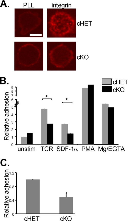
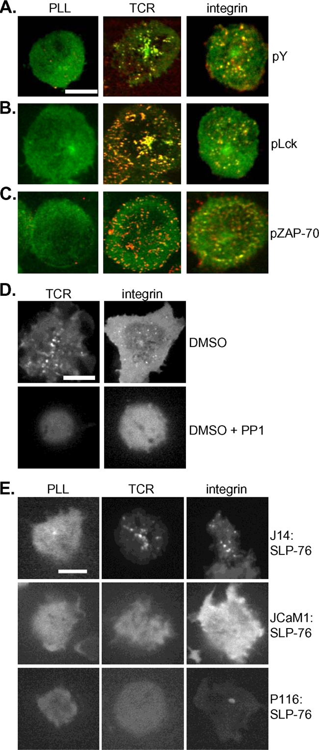
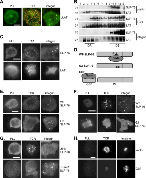
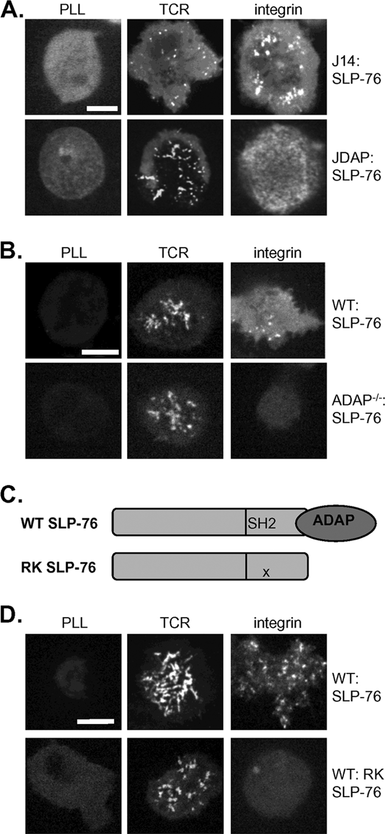
The adapter protein SH2 domain-containing leukocyte protein of 76 kDa (SLP-76) is an essential mediator of signaling from the T-cell antigen receptor (TCR). We report here that SLP-76 also mediates signaling downstream of integrins in T cells and that SLP-76-deficient T cells fail to support adhesion to integrin ligands. In response to both TCR and integrin stimulation, SLP-76 relocalizes to surface microclusters that colocalize with phosphorylated signaling proteins. Disruption of SLP-76 recruitment to the protein named LAT (linker for activation of T cells) inhibits SLP-76 clustering downstream of the TCR but not downstream of integrins. Conversely, an SLP-76 mutant unable to bind ADAP (adhesion and degranulation-promoting adapter protein) forms clusters following TCR but not integrin engagement and fails to support T-cell adhesion to integrin ligands. These findings demonstrate that SLP-76 relocalizes to integrin-initiated signaling complexes by a mechanism different from that employed during TCR signaling and that SLP-76 relocalization corresponds to SLP-76-dependent integrin function in T cells.


Figures

FIG. 1.
FIG. 1.
SLP-76 is dispensable for signaling from the chemokine receptor CXCR4. (A) Temporally controlled deletion of SLP-76 was accomplished through mice expressing a SLP-76 allele flanked with loxP restriction sites by treatment with tamoxifen. Following excision of one floxed SLP-76 allele (loxP-flanked box), cHET mice posressed one remaining copy of SLP-76 (top) and cells from cKO mice possessed no remaining SLP-76 allele (bottom). (B) SLP-76 cHET and cKO cells were allowed to migrate across a transwell barrier into medium alone (left panel) or into a source of SDF-1α (right panel), and results are presented as mean numbers of recovered cells (103) ± standard deviations determined in six independent experiments. (C) SLP-76 cHET and cKO T cells were left unstimulated (unstim) or were stimulated with 1 ng of SDF-1α/ml for 2 and 10 min, and active Rap1 was precipitated from cell lysates. Rap1 activity levels were similarly upregulated in SLP-76 cHET and cKO cells. (D) SLP-76 cHET and cKO T cells were left unstimulated or were stimulated with SDF-1α, and binding of soluble mICAM-1 was evaluated by flow cytometry as a measurement of LFA-1 affinity. cHET and cKO cells bind equivalent levels of ICAM-1 in response to SDF-1α stimulation. Max, maximum; iso, isotype.
FIG. 2.
FIG. 2.
SLP-76 is required for T-cell integrin signaling and adhesion to ICAM-1. (A) Proximal signaling from integrins is defective in SLP-76 cKO cells, as measured by immunofluorescence microscopy. LFA-1 on SLP-76 cHET and cKO T cells was stimulated by pretreatment with SDF-1α followed by deposition onto the ligand ICAM-1. Following 10 min of stimulation, cells were fixed and stained for total phosphotyrosine. Clusters of phosphotyrosine staining appear in cHET but not in cKO cells. Bar, 5 μm. (B) Adhesion of SLP-76 cHET and cKO cells to ICAM-1 was measured among cells subjected to the indicated stimuli. Adhesion levels ranged from 5 to 10% in unstimulated (unstim) cells to 80% at maximum. Data were normalized to unstimulated cHET cell results and represent means ± standard deviations of the results of 10 independent experiments. Asterisks indicate a statistical significance of P < 0.05. (C) Levels of adhesion of SLP-76 cKO and cHET cells to SDF-1α and ICAM-1 under flow conditions were compared. Data represent means ± standard deviations normalized to unstimulated cHET cell results obtained in three independent experiments.
FIG. 3.
FIG. 3.
SLP-76 relocalizes to microclusters at the cell surface in response to TCR and integrin stimulation. (A) J14:GFP-SLP-76 T cells were deposited onto PLL, TCR, and integrin (ICAM-1)-stimulating surfaces, and recruitment of GFP-SLP to microclusters at the contact site is shown. Bars, 5 μm. (B and C) GFP-SLP-76 was recruited to microclusters in primary T-cell blasts (B) and naïve murine T cells (C). Image results are representative of at least five independent experiments.
FIG. 4.
FIG. 4.
PTKs direct SLP-76 relocalization to integrin-initiated microclusters. (A) J14 cells expressing GFP-SLP-76 (green) were deposited onto unstimulatory or TCR or integrin (FN) stimulatory surfaces, fixed, and stained for total phosphorylated tyrosine (pY) (red); merged images are shown. Bars, 5 μm. (B) GFP-SLP-76 (green) colocalized with phospho-Lck (pLck) (red). (C) GFP-SLP-76 (green) colocalized with phospho-ZAP-70 (pZAP-70) (red). (D) J14:GFP-SLP-76 cells were preincubated with either dimethyl sulfoxide (DMSO) carrier alone or DMSO plus PP1 and were stimulated by TCR or integrin (FN). (E) JCaM1 and P116 Jurkat T cells transfected with GFP-SLP-76 were deposited onto TCR and integrin (FN) stimulatory surfaces; the resulting GFP-SLP-76 clusters are shown. All image results are representative of at least three independent experiments.
FIG. 5.
FIG. 5.
TCR and integrin-initiated SLP-76 microclusters exhibit different dynamic properties. (A) J14:GFP-SLP-76 cells were deposited onto TCR and integrin (FN) stimulatory surfaces and imaged over time by TIRF microscopy, and snapshots from indicated time points (indicated in seconds) are shown. Bars, 5 μm. (B) GFP-SLP-76 clusters were tracked at all time points, and trajectories are shown overlaid on the sample cell. (C) (Left panel) The displacement of GFP-SLP-76 clusters was calculated, and the results are shown with respect to elapsed time, with TCR results represented in black and integrin (FN) results in gray. (Right panel) Numbers of GFP-SLP-76 clusters in representative cells were counted at multiple time points, and percentages representing the maximum numbers of clusters counted at any time point are shown. Data represent means ± standard deviations of the results of 10 TCR and 4 FN experiments.
FIG. 6.
FIG. 6.
SLP-76 relocalization downstream of integrins is independent of LAT. (A) J14 cells expressing GFP-SLP-76 (green) were deposited onto unstimulatory (unstim) and TCR and integrin (FN) stimulatory surfaces, fixed, and stained for phospho-LAT Y-191 (red); color-merged images are shown. Bars, 5 μm. (B) J14:GFP-SLP-76 cells were stimulated by TCR and integrins (FN), and density ultracentrifugation was used to separate detergent-resistant (DR) (lanes 2 to 5) from detergent-soluble (DS) (lanes 9 to 12) protein fractions, which were then subjected to immunoblot analysis. (C) LAT−/− JCaM2 Jurkat cells were transfected with GFP-LAT and stimulated by TCR and integrins (FN). (D) The G2 SLP-76 mutant was unable to support Gads binding or Gads-mediated recruitment to LAT, while the GBF competed with SLP-76 for Gads binding. (E and F) J14 (E) and primary (F) T-cell blasts expressing GFP-G2 and WT SLP-76 were stimulated by TCR and integrin (FN and ICAM-1, respectively). (G) JCaM2 T cells supported the formation of integrin (FN)-initiated but not TCR-initiated GFP-SLP-76 microclusters. (H) Primary T-cell blasts transduced with both GFP-WT SLP-76 and either vector alone or GBF peptide were stimulated by TCR or integrins (ICAM-1). All image results are representative of at least five independent experiments.
FIG. 7.
FIG. 7.
SLP-76 relocalization downstream of integrins requires ADAP. (A and B) JDAP Jurkat T cells (A) and ADAP−/− primary T-cell blasts (B) transduced with GFP-SLP-76 were stimulated by TCR and integrin (FN and ICAM-1, respectively). Bars, 5 μm. (C) The RK mutation in the SH2 domain of SLP-76 disrupts SLP-76 association with ADAP. (D) Primary T-cell blasts were transduced with WT or RK-SLP-76 and stimulated by TCR or integrins (ICAM-1). All image results are representative of at least five independent experiments.
FIG. 8.
FIG. 8.
SLP-76 relocalization correlates with support of integrin function. (A) Adhesion to ICAM-1 among naïve CD4-Cre SLP-76 cHET, cKO, cKO:RK SLP-76, or ADAP−/− T cells was measured in response to the indicated stimuli. Adhesion levels ranged from 5 to 10% in unstimulated (unstim) cells to 80% at maximum. Data represent means ± standard deviations normalized to unstimulated cHET cell results from four experiments. Asterisks indicate a statistical significance of P < 0.05. (B) Adhesion to ICAM-1 was measured among Ubc-CreT2 SLP-76 cKO T-cell blasts retrovirally reconstituted with vector only or with WT, G2, or SLP-76. Data represent means ± standard deviations normalized to unstimulated WT cell results from three experiments. ns, not significant.

References

    1. Abraham, R. T., and A. Weiss. 2004. Jurkat T cells and development of the T-cell receptor signalling paradigm. Nat. Rev. Immunol. 4:301-308. - PubMed
    1. Arias-Salgado, E. G., S. Lizano, S. Sarkar, J. S. Brugge, M. H. Ginsberg, and S. J. Shattil. 2003. Src kinase activation by direct interaction with the integrin beta cytoplasmic domain. Proc. Natl. Acad. Sci. USA 100:13298-13302. - PMC - PubMed
    1. Baker, R. G., and G. A. Koretzky. 2008. Regulation of T cell integrin function by adapter proteins. Immunol. Res. 42:132-144. - PubMed
    1. Balagopalan, L., V. A. Barr, C. L. Sommers, M. Barda-Saad, A. Goyal, M. S. Isakowitz, and L. E. Samelson. 2007. c-Cbl-mediated regulation of LAT-nucleated signaling complexes. Mol. Cell. Biol. 27:8622-8636. - PMC - PubMed
    1. Barr, V. A., L. Balagopalan, M. Barda-Saad, R. Polishchuk, H. Boukari, S. C. Bunnell, K. M. Bernot, Y. Toda, R. Nossal, and L. E. Samelson. 2006. T-cell antigen receptor-induced signaling complexes: internalization via a cholesterol-dependent endocytic pathway. Traffic 7:1143-1162. - PubMed

Publication types

MeSH terms