. 2009 Aug 10;186(3):437-46.
doi: 10.1083/jcb.200903082.

Asymmetry of inner dynein arms and inter-doublet links in Chlamydomonas flagella

Affiliations

Asymmetry of inner dynein arms and inter-doublet links in Chlamydomonas flagella

Khanh Huy Bui et al. J Cell Biol. .

Abstract

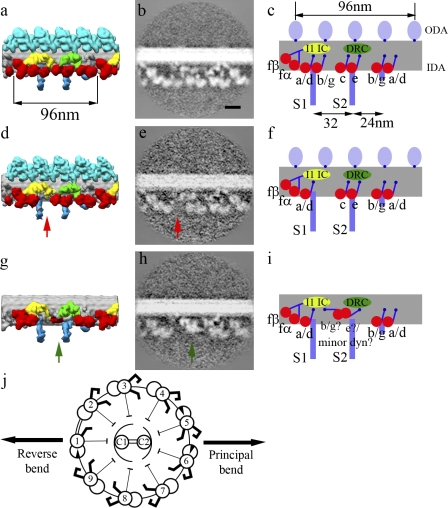
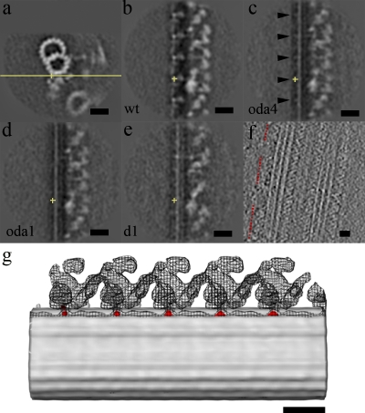
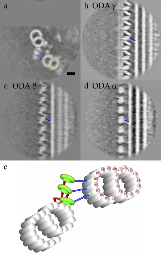
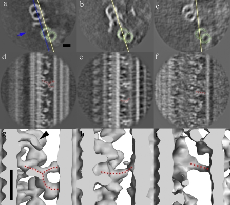
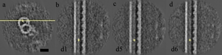
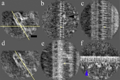
Although the widely shared "9 + 2" structure of axonemes is thought to be highly symmetrical, axonemes show asymmetrical bending during planar and conical motion. In this study, using electron cryotomography and single particle averaging, we demonstrate an asymmetrical molecular arrangement of proteins binding to the nine microtubule doublets in Chlamydomonas reinhardtii flagella. The eight inner arm dynein heavy chains regulate and determine flagellar waveform. Among these, one heavy chain (dynein c) is missing on one microtubule doublet (this doublet also lacks the outer dynein arm), and another dynein heavy chain (dynein b or g) is missing on the adjacent doublet. Some dynein heavy chains either show an abnormal conformation or were replaced by other proteins, possibly minor dyneins. In addition to nexin, there are two additional linkages between specific pairs of doublets. Interestingly, all these exceptional arrangements take place on doublets on opposite sides of the axoneme, suggesting that the transverse functional asymmetry of the axoneme causes an in-plane bending motion.


Figures

Figure 1.
Figure 1.
The architecture of the IDAs on individual microtubule doublets. (a, d, and g) The surface rendering of doublets 2–8 (a), 9 (d), and 1 (g). (b, c, e, f, h, and i) Same section across the averaged density map and the schematic representation of doublets 2–8 (b and c), 9 (e and f), and 1 (h and i) showing the inner arm dynein pattern. Red arrows show the position of the absent dynein b/g adjacent to radial spoke S1, and green arrows show the bulb density on doublet 1. Red, IDA; light blue, ODA; blue, radial spokes; green, DRC; yellow, light chain (LC)/IC of dynein f. The heads of radial spokes were also seen (not depicted) but have been trimmed in these figures to show only the area around the doublets. (j) The doublet-numbering order was performed as described by Hoops and Witman (1983), with doublets 1, 5, and 6 having the beaklike projection and a 1-2 bridge between doublets 1 and 2. Double-ended black arrows indicate the beating plane. Bar, 20 nm.
Figure 2.
Figure 2.
Comparison of doublet 1 and the common IDA architecture. (a) Overall view and 90° view. (b–d) Enlarged images of doublet 1 (boxed regions in a) shown at different viewing angles, with the common architecture (average doublets 2–8) used as a control on the right. Blue arrowheads, folded tail of dynein e; black arrowheads, positions where the dynein c tail should emerge (Bui et al., 2008) but is missing on doublet 1; orange arrowheads, additional density on doublet 1 behind dynein e. (e) Sections of the average of doublet 1 (d1; left) and doublets 2–8 (dc; right) of mutant ida4, showing that the lack of dynein c does not change the appearance of the bulb density on doublet 1. The green arrow shows the bulb density on doublet 1. Bar, 20 nm.
Figure 3.
Figure 3.
DC analysis on wild type, oda4 (lacking ODA but retaining DC), oda1 (lacking both ODA and DC), and the doublet 1 average from wild type, oda11, oda4, and oda4s7. The yellow crosses show the center of the extra density and are at the same position in all cross sections. (a) Cross section of the common IDA architecture of wild type with the yellow line indicating the plane of the yz section. (b) yz section of wild type (wt) showing the 24-nm repeat. (c) yz section of oda4 also showing the 24-nm spacing (black arrowheads). (d) No 24-nm repeat is shown in oda1. (e) yz section of the doublet 1 average. (f) Horizontal section of a tomogram involving doublet 1 from wild-type flagellum. ODA is present but with no regular 24-nm periodicity. Red dotted lines indicate the regions with ODA. (g) Overlap of surface rendering of wild type, oda4, and the difference map between oda4 and oda1, showing that the 24-nm periodicity from the difference map (red) is at exactly the same place as the interface of ODA with the A-tubule. The surface rendering of wild type and oda4 are represented by mesh and gray surfaces, respectively. Bars, 20 nm.
Figure 4.
Figure 4.
The attachment of the ODA stalk to the adjacent microtubule. (a–d) It is shown as a cross section (a), ODA γ (b), ODA β (c), and ODA α (d). The dotted blue lines indicate the stalk. The green circles indicate the protofilaments in the adjacent B-tubule. (e) A model of ODA stalk interaction with the adjacent B-tubule is shown. Bar, 20 nm.
Figure 5.
Figure 5.
The nexin linker, IDL2, and IDL3. (a–i) Cross section and tilted views (yellow lines) and surface rendering, showing the circumferential nexin in all the doublets (a, d, and g), IDL2 from doublets 4, 5, and 9 (b, e, and h), and IDL3 from doublet 1 (c, f, and i). The green circles indicate the protofilaments in the adjacent B-tubule. The dotted red lines are drawn along the nexin, IDL2, and IDL3. The blue line and arrow in a indicate the cross section in the surface rendering (g–i) and the direction of view, respectively. The black arrowhead in g shows the protrusion above the radial spoke S3-like feature, which was identified as part of the nexin in our previous study (Bui et al., 2008), but it is now clear that it is not connecting to the nexin. Bars, 20 nm.
Figure 6.
Figure 6.
The beaklike projections inside doublets. (a) The vertical section shows the plane of view of the cross sections (yellow line). (b–d) Longitudinal sections of doublets 1 (b), 5 (c), and 6 (d). The yellow crosses in the yz cross sections show the beaklike feature and are at the same position as the yellow cross in a. Bar, 20 nm.
Figure 7.
Figure 7.
Linkage between doublets 1 and 2 when the 1-2 bridge is present in a cross section. (a, c, and e) The 1-2 bridge (8-nm period) in the xy plane (a), xz plane (c), and tilted section along the linkage (e). (b and d) The other two dominant linkages in the cross section (b) and tilted section along the two dominant linkages (d) are shown. The yellow crosses in b and c and e indicate the same position as the yellow crosses in a and d, respectively. Yellow lines in a and d indicate the planes of sections in b and c and e, respectively. The black arrowhead in b indicates the region where the inner bridge is discontinued. (f) Longitudinal section showing IDA of wild-type doublet 1 in the region of the 1-2 bridge. The blue arrowhead points to the difference from doublet 1 structure outside the 1-2 bridge region (Fig. 1 h). Bar, 20 nm.
Figure 8.
Figure 8.
Hypothesis of the mechanism of planar asymmetrical bending motion of flagella. (a) Doublet order of flagella at the basal body of C. reinhardtii (Hoops and Witman, 1983), showing that doublets 1 of the two flagella are facing each other. Arrows indicate the plane of bend. (b) Illustration of the generation of planar asymmetric waveform by strong and weak sides is shown.

References

    1. Adams G.M., Huang B., Piperno G., Luck D.J. 1981. Central-pair microtubular complex of Chlamydomonas flagella: polypeptide composition as revealed by analysis of mutants.J. Cell Biol. 91:69–76 - PMC - PubMed
    1. Afzelius B. 1959. Electron microscopy of the sperm tail; results obtained with a new fixative.J. Biophys. Biochem. Cytol. 5:269–278 - PMC - PubMed
    1. Bannai H., Yoshimura M., Takahashi K., Shingyoji C. 2000. Calcium regulation of microtubule sliding in reactivated sea urchin sperm flagella.J. Cell Sci. 113:831–839 - PubMed
    1. Bessen M., Fay R.B., Witman G.B. 1980. Calcium control of waveform in isolated flagellar axonemes of Chlamydomonas.J. Cell Biol. 86:446–455 - PMC - PubMed
    1. Bozkurt H.H., Woolley D.M. 1993. Morphology of nexin links in relation to interdoublet sliding in the sperm flagellum.Cell Motil. Cytoskeleton. 24:109–118 - PubMed

Publication types

MeSH terms