. 2009 Aug 25;106(34):14564-9.
doi: 10.1073/pnas.0813319106. Epub 2009 Aug 10.

Autophagy genes protect against Salmonella typhimurium infection and mediate insulin signaling-regulated pathogen resistance

Affiliations

Autophagy genes protect against Salmonella typhimurium infection and mediate insulin signaling-regulated pathogen resistance

Kailiang Jia et al. Proc Natl Acad Sci U S A. .

Abstract

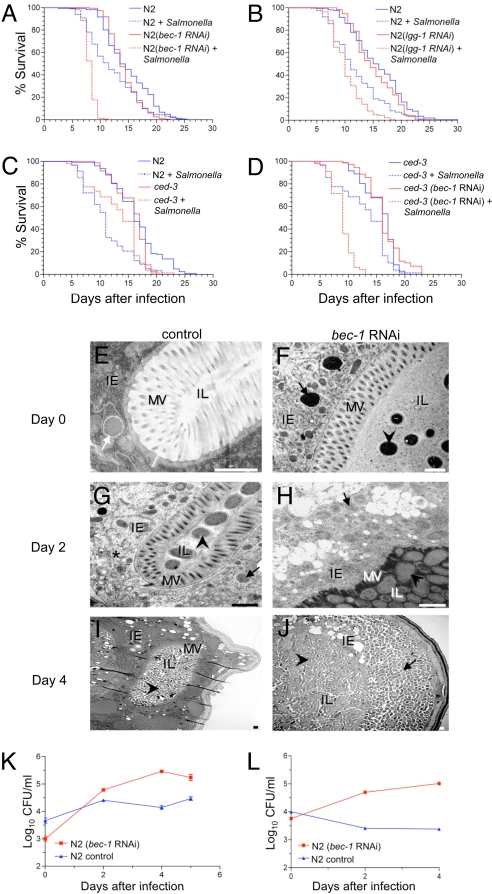
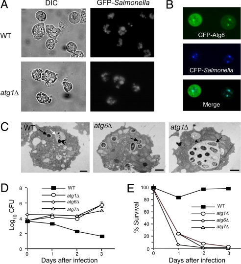
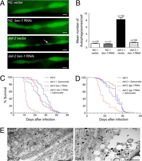
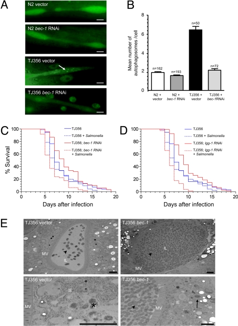
A conserved insulin-like pathway modulates both aging and pathogen resistance in Caenorhabditis elegans. However, the specific innate effector functions that mediate this pathogen resistance are largely unknown. Autophagy, a lysosomal degradation pathway, plays a role in controlling intracellular bacterial pathogen infections in cultured cells, but less is known about its role at the organismal level. We examined the effects of autophagy gene inactivation on Salmonella enterica Serovar Typhimurium (Salmonella typhimurium) infection in 2 model organisms, Caenorhabditis elegans and Dictyostelium discoideum. In both organisms, genetic inactivation of the autophagy pathway increases bacterial intracellular replication, decreases animal lifespan, and results in apoptotic-independent death. In C. elegans, genetic knockdown of autophagy genes abrogates pathogen resistance conferred by a loss-of-function mutation, daf-2(e1370), in the insulin-like tyrosine kinase receptor or by over-expression of the DAF-16 FOXO transcription factor. Thus, autophagy genes play an essential role in host defense in vivo against an intracellular bacterial pathogen and mediate pathogen resistance in long-lived mutant nematodes.


Conflict of interest statement

The authors declare no conflict of interest.

Figures

Fig. 1.
Fig. 1.
Atg genes mediate host defense against Salmonella in C. elegans. (A and B) Survival curves of wild-type (N2) animals treated with either control vector or indicated atg gene RNAi-feeding plasmids following a 2-day exposure to S. typhimurium or normal food (i.e., nonpathogenic Escherichia coli) at 20 °C (see SI Materials and Methods for details). (C and D) Survival curves of wild-type (N2) and ced-3(n717)-mutant animals following a 2-day exposure to S. typhimurium or normal food without atg gene RNAi treatment (C) or with control vector or bec-1 RNAi treatment (D) at 20 °C. For (A) to (D), see Table S1 for statistical details. (E–J) Representative EMs of Salmonella-infected control N2 animals and bec-1-RNAi animals at day 0 (E and F), 2 (G and H), and 4 (I and J) after a 2-day Salmonella ingestion period. (F, G, I, and J) Arrowheads denote intraluminal bacteria. (E) White arrow denotes bacterial-containing early autophagosome. (G) Asterisk denotes autolysosome with partially degraded bacterial debris. (F–H and J) Black arrows denote SCVs inside intestinal epithelial cells. IE, intestinal epithelial cells; IL, intestinal lumen; MV, microvilli. (Scale bars, 1 μm.) (K and L) Growth curves of S. typhimurium in N2 animals treated with either control vector or bec-1-RNAi feeding plasmids in the absence (K) or presence (L) of 100 μg/ml gentamicin. For (K) and (L), values represent mean ± SEM for triplicate samples of ≈10 animals per treatment group per sample. Similar results were observed in two independent experiments.
Fig. 2.
Fig. 2.
Atg genes restrict intracellular Salmonella replication and pathogenicity in D. discoideum. (A) Internalization and vacuolar localization of GFP-Salmonella in both wild-type (WT) and atg1 mutant Dictyostelium at 2-h postinfection (p.i.). Similar results were observed in atg6Δ and atg7Δ Dictyostelium (data not shown). (B) Colocalization of CFP-Salmonella and a transgenic autophagosomal marker, GFP-Atg8, at 2-h p.i. in wild-type Dictyostelium. (C) Representative EMs 12-h p.i. of wild-type Dictyostelium showing an intact SCV (black arrow) and an autolysosome (white arrow) containing a partially degraded Salmonella bacterieum (arrowhead), and of atg1Δ and atg6Δ Dictyostelium that lack autolyosomes but have SCVs (black arrows) that contain multiple organisms, indicative of active intracellular bacterial multiplication. (Scale bars, 1 μm.) (D) Growth of intracellular Salmonella in wild-type and atg gene-mutant Dictyostelium strains. (E) Survival of Dictyostelium infected in (D). For (D) and (E), results represent mean ± SEM for triplicate samples and similar results were observed in three independent experiments.
Fig. 3.
Fig. 3.
Atg genes mediate insulin signaling-regulated resistance against Salmonella in C. elegans. (A) Representative images of autophagosomes (GFP::LGG-1 dots) in seam cells of N2 wild-type and daf-2-mutant animals treated either with RNAi control vector or bec-1 RNAi. The arrow denotes a representative autophagosome. (Scale bars, 2 μM.) (B) Quantification of autophagosomes per seam cell (mean ± SEM) for each genotype. n = number of seam cells per group in ≈20 animals. Similar results were obtained in two independent experiments. (C and D) Survival curves of daf-2(e1370)-mutant animals treated with either control or indicated atg gene-RNAi feeding plasmids following a 2-day exposure to S. typhimurium or normal food at 20 °C. See Table S1 for statistical details. (E) Representative EMs of Salmonella-infected control and bec-1-RNAi daf-2(e1370) animals two days after a 2-day Salmonella ingestion period. The asterisk denotes a representative autolysosome containing partially degraded bacteria. The arrow denotes representative SCV. (Scale bars, 2 μM.)
Fig. 4.
Fig. 4.
Atg genes are required for DAF-16-mediated resistance to Salmonella infection. (A) Representative pictures of autophagosomes in seam cells of N2 wild-type and TJ356 animals treated either with RNAi control vector or bec-1 RNAi. TJ356 is heterozygous for both gfp::lgg-1 and daf-16::gfp. The oval-shaped green staining near the seam cells in TJ356 animals represent nuclei of intestinal cells that express DAF-16::GFP. The arrow denotes a representative autophagosome. (Scale bars, 2 μM.) (B) Quantification of autophagosomes per seam cell (mean ± SEM) for each genotype. n = number of seam cells per group in ≈20 animals. Similar results were obtained in two independent experiments. (C and D) Survival curves of TJ356 animals treated with either control or indicated atg gene RNAi-feeding plasmids following exposure to S. typhimurium or normal food at 25 °C. n = number of animals per treatment group. See Table S1 for statistical details. (E) Representative EMs of Salmonella-infected control and bec-1-RNAi TJ356 animals two days after a 2-day Salmonella ingestion period. The asterisk denotes a representative autolysosome containing partially degraded bacteria. Arrowheads represent intestinal luminal bacteria and the arrow denotes representative SCV-containing bacteria. (Scale bars, 2 μM.)

References

    1. Gavazzi G, Krause KH. Ageing and infection. Lancet Infect Dis. 2002;2:659–666. - PubMed
    1. Kenyon C. The plasticity of aging: Insights from long-lived mutants. Cell. 2005;120:449–460. - PubMed
    1. Garsin D, et al. Long-lived C. elegans daf-2 mutants are resistant to bacterial pathogens. Science. 2003;300:1921. - PubMed
    1. Levine B, Deretic V. Unveiling the roles of autophagy in innate and adaptive immunity. Nat Rev Immunol. 2007;7:767–777. - PMC - PubMed
    1. Melendez A, et al. Autophagy genes are essential for dauer development and lifespan extension in C. elegans. Science. 2003;301:1387–1391. - PubMed

Publication types

MeSH terms

LinkOut - more resources