. 2009 Aug 18;106(33):13915-20.
doi: 10.1073/pnas.0905780106. Epub 2009 Aug 10.

Triplet-repeat oligonucleotide-mediated reversal of RNA toxicity in myotonic dystrophy

Affiliations

Triplet-repeat oligonucleotide-mediated reversal of RNA toxicity in myotonic dystrophy

Susan A M Mulders et al. Proc Natl Acad Sci U S A. .

Abstract

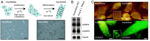
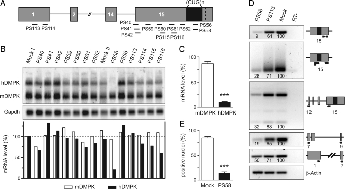
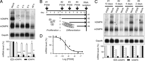
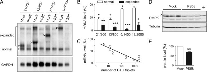
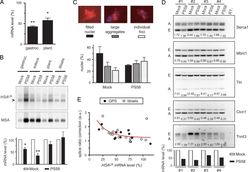
Myotonic dystrophy type 1 (DM1) is caused by toxicity of an expanded, noncoding (CUG)n tract in DM protein kinase (DMPK) transcripts. According to current evidence the long (CUG)n segment is involved in entrapment of muscleblind (Mbnl) proteins in ribonuclear aggregates and stabilized expression of CUG binding protein 1 (CUGBP1), causing aberrant premRNA splicing and associated pathogenesis in DM1 patients. Here, we report on the use of antisense oligonucleotides (AONs) in a therapeutic strategy for reversal of RNA-gain-of-function toxicity. Using a previously undescribed mouse DM1 myoblast-myotube cell model and DM1 patient cells as screening tools, we have identified a fully 2'-O-methyl-phosphorothioate-modified (CAG)7 AON that silences mutant DMPK RNA expression and reduces the number of ribonuclear aggregates in a selective and (CUG)n-length-dependent manner. Direct administration of this AON in muscle of DM1 mouse models in vivo caused a significant reduction in the level of toxic (CUG)n RNA and a normalizing effect on aberrant premRNA splicing. Our data demonstrate proof of principle for therapeutic use of simple sequence AONs in DM1 and potentially other unstable microsatellite diseases.


Conflict of interest statement

Conflict of interest: P.v.K.-R., S.J.d.K., and G.J.P. report being employed by or having an equity interest in Prosensa B.V. The method described in this paper is the subject of a patent application (inventors S.J.d.K, G.J.P., and D.G.W.).

Figures

Fig. 1.
Fig. 1.
Novel cell model for testing DM1 therapy. (A) Conditionally immortalized myoblasts derived from DM500 mice differentiate to myotubes under low serum conditions. Bars, 30 μm. (B) Northern blot analysis of RNA isolated by 2 different methods from DM500 myoblasts demonstrated expression of transgenic hDMPK mRNA bearing a (CUG)500 tract next to endogenous mDMPK mRNA. (C) FISH using a (CAG)7 probe revealed ribonuclear (CUG)n foci, mainly in cell nuclei. A (CUG)7 probe was used as negative control. Insets show nuclei at higher magnification. Bars, 10 μm.
Fig. 2.
Fig. 2.
PS58-mediated silencing of expanded hDMPK transcripts in DM500 cells. (A) Location of target sites of AONs (black bars) along the hDMPK premRNA. (B) Northern blot analysis to detect ability of AONs to silence hDMPK mRNA in DM500 myotubes. Gapdh was used as loading control. Oligos were tested at least twice; a representative blot is shown. (C) PS58, directed at the (CUG)n segment, was the most successful AON (P < 0.001, n = 19). (D) PS58 activity was corroborated by semiquantitative RT-PCR analysis of individual segments of the hDMPK transcript (primers indicated with black arrows). Numerals in lanes indicate relative abundance to mock samples (set at 100), using β-actin for normalization. A representative result from 2 experiments is shown. (E) A reduction in the number of nuclei containing (CUG)n foci was observed after PS58 treatment of DM500 myoblasts (P < 0.001, n = 4).
Fig. 3.
Fig. 3.
PS58-induced silencing is rapid, persistent, and occurs with high efficacy. (A) DM500 myotubes were mock-treated or treated with PS58. RNA was isolated 2–48 h after start of transfection. Northern blot analysis indicated that expanded hDMPK was degraded within 2 hours. A representative blot is shown. The means of 3 experiments are summarized in the Bottom panel (P < 0.05). (B and C) DM500 myoblasts and myotubes in different stages of myogenesis were treated with a single 200-nM PS58 dose or mock-treated and then cultured for up to 10 days. Northern blot analysis demonstrated that hDMPK RNA remained undetectable for up to 10 days. (D) Concentration-response curve in DM500 myotubes. Data points are the mean of at least 3 measurements.
Fig. 4.
Fig. 4.
PS58 silences preferentially expanded DMPK transcripts in patient cells. (A) DM1 myoblasts expressing different expanded and normal-sized DMPK alleles were treated with PS58 or mock treated. Northern blot analysis indicated that expanded mRNA was strongly reduced whereas normal-sized DMPK transcripts were less sensitive to breakdown. A representative blot is shown. (B) Quantification of 3 experiments as described in A (P < 0.05). (C) A clear correlation was demonstrated between the number of CTG triplets and PS58 efficacy (P < 0.05, Spearman correlation, r = −0.79). (D and E) Myoblasts (21/200) were analyzed by Western blotting 72 h after mock or PS58 treatment. A muscle sample from a DMPK KO mouse (−/−) served as negative control. A 29% reduction in DMPK protein (arrowhead) was observed (P < 0.01, n = 3).
Fig. 5.
Fig. 5.
PS58 reverses molecular features in DM1 mouse models in vivo. (A) PS58 was injected in the GPS complex of DM500 mice, followed by RNA isolation at day 15. Semiquantitative RT-PCR analysis demonstrated hDMPK mRNA silencing in gastrocnemius (P < 0.01, n = 3) and plantaris (P < 0.05, n = 3) (see Fig. S5). (B) A similar protocol was used in HSAlr mice. TA muscle served as uninjected control. HSAlr transcripts were quantified on Northern blot, using mouse α-actin (MSA) for normalization. The blot represents 1 of 4 independent experiments. The histogram shows average values: lower HSAlr mRNA levels were observed in soleus (35 ± 8%; P < 0.01) and gastrocnemius (61 ± 7%; P = 0.01), but not in plantaris (81 ± 11%; P = 0.19). (C) FISH analysis was performed on sections of PS58- or mock-injected HSAlr gastrocnemius muscle. (CUG)n foci were found in every nucleus. Nuclei were categorized in 3 groups according to their (CUG)n foci appearance (Top panels each show 1 representative nucleus). A significant shift toward smaller and fewer foci was observed after PS58 treatment (P < 0.01, n = 2). (D) TA muscle in HSAlr mice was electroporated with PS58 or mock treated. RNA was isolated after 7 days. Northern blotting showed significantly lower HSAlr mRNA levels after PS58 treatment (Bottom panel; 51 ± 7%; P < 0.01, n = 4). Splice modes were investigated by RT-PCR. Numerals indicate embryonic (E):adult (A) splice ratio in each lane. Lower ratios in PS58-treated samples, compared to their mock-treated samples, demonstrate positive effects on alternative splicing (see also Fig. S7). (E) Correlation between splicing correction and HSAlr mRNA levels in PS58-treated muscles. Each symbol represents a TA muscle or 1 muscle from the GPS complex. Splice ratio correction illustrates the mean effect on alternative splicing of 5 transcripts (see D) (P < 0.01; Spearman correlation, r = −0.64; n = 27).

References

    1. Groenen P, Wieringa B. Expanding complexity in myotonic dystrophy. BioEssays. 1998;20:901–912. - PubMed
    1. Wheeler TM, Thornton CA. Myotonic dystrophy: RNA-mediated muscle disease. Curr Opin Neurol. 2007;20:572–576. - PubMed
    1. Miller JW, et al. Recruitment of human muscleblind proteins to (CUG)(n) expansions associated with myotonic dystrophy. EMBO J. 2000;19:4439–4448. - PMC - PubMed
    1. Kuyumcu-Martinez NM, Wang GS, Cooper TA. Increased steady-state levels of CUGBP1 in myotonic dystrophy 1 are due to PKC-mediated hyperphosphorylation. Mol Cell. 2007;28:68–78. - PMC - PubMed
    1. Mankodi A, et al. Myotonic dystrophy in transgenic mice expressing an expanded CUG repeat. Science. 2000;289:1769–1772. - PubMed

Publication types