. 2009 Aug 18;106(33):14028-33.
doi: 10.1073/pnas.0906796106. Epub 2009 Aug 10.

A role for the TGFbeta-Par6 polarity pathway in breast cancer progression

Affiliations

A role for the TGFbeta-Par6 polarity pathway in breast cancer progression

Alicia M Viloria-Petit et al. Proc Natl Acad Sci U S A. .

Abstract

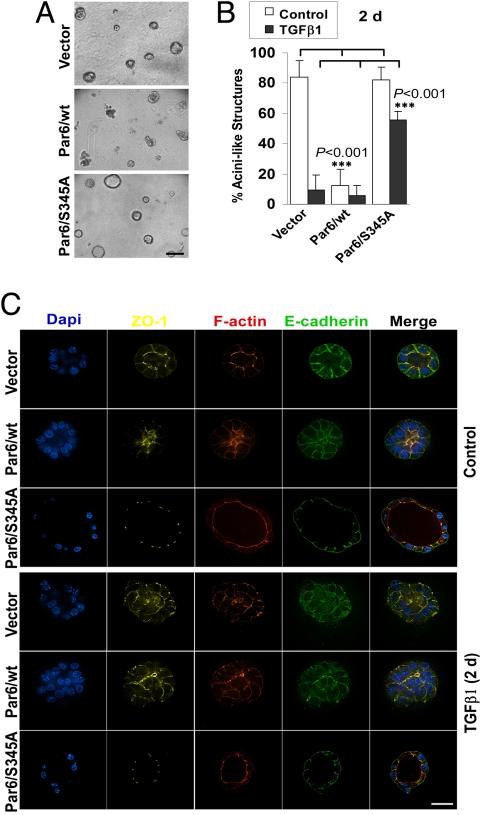
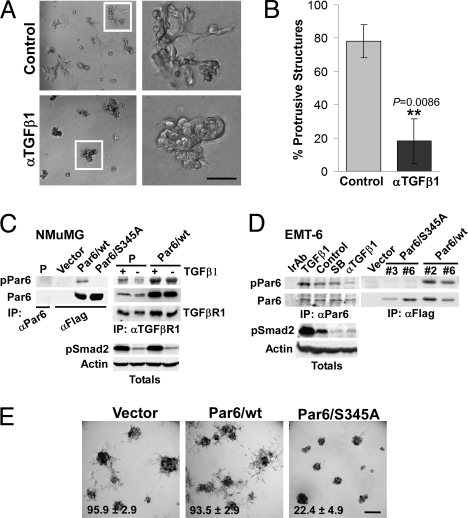
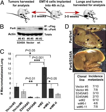
The role of polarity signaling in cancer metastasis is ill defined. Using two three-dimensional culture models of mammary epithelial cells and an orthotopic mouse model of breast cancer, we reveal that Par6 signaling, which is regulated directly by TGFbeta, plays a role in breast cancer metastasis. Interference with Par6 signaling blocked TGFbeta-dependent loss of polarity in acini-like structures formed by non-transformed mammary cells grown in three-dimensional structures and suppressed the protrusive morphology of mesenchymal-like invasive mammary tumor cells without rescuing E-cadherin expression. Moreover, blockade of Par6 signaling in an in vivo orthotopic model of metastatic breast cancer induced the formation of ZO-1-positive epithelium-like structures in the primary tumor and suppressed metastasis to the lungs. Analysis of the pathway in tissue microarrays of human breast tumors further revealed that Par6 activation correlated with markers of the basal carcinoma subtype in BRCA1-associated tumors. These studies thus reveal a key role for polarity signaling and the control of morphologic transformation in breast cancer metastasis.


Conflict of interest statement

The authors declare no conflict of interest.

Figures

Fig. 1.
Fig. 1.
Activation of the TGFβ-Par6 pathway interferes with the formation of polarized acini-like structures by NMuMG cells. (A) Gross morphology of 9-day-old 3D cultures of NMuMG lines expressing empty vector (Vector), wt, or S345A Par6. (B) Quantification of acini-like structures. Nine-day-old structures were treated with TGFβ (500 pM; black bars) or without (Control; white bars) for 2 days (2d) and the percentage of acini-like structures (containing a lumen) quantified. Most Par6/wt structures lacked a lumen under basal conditions and maintained their abnormal morphology after TGFβ treatment. In sharp contrast, about 60% of Par6/S345A structures remain polarized after TGFβ exposure. (C) The TGFβ-Par6 pathway disrupts polarity in NMuMG 3D structures. TGFβ treated or untreated structures as in B were immunostained for nuclei (DAPI, blue) and polarity markers, followed by confocal microscopy analysis. Untreated vector and Par6/S345A structures (Top) had well-defined lumens, with ZO-1 (yellow) and F-actin (red) localized to the apical, TJ region, and E-cadherin (green) localized to the AJ, basal to ZO-1. Par6/wt structures had disorganized ZO-1 and F-actin and were lumenless. TGFβ treatment (Bottom) caused ZO-1, F-actin, and E-cadherin mislocalization in both Vector and Par6/wt-expressing structures, but not in S345A structures. (Scale bar in A, 100 μm; C, 20 μm.)
Fig. 2.
Fig. 2.
Autocrine TGFβ signaling regulates the protrusive, mesenchymal phenotype of EMT6 cells via the Par6 pathway. (A and B) EMT6 cells were grown in Matrigel for 5 days and continuously treated with an anti-TGFβ1 Ab (αTGFβ1) at 10 μg/mL or left untreated, as indicated. Bright field images of 3D structures are shown in A, while quantification of protrusive structures formed in control or αTGFβ1-treated cultures is shown in B. (C) Characterization of a Par6S345P (pPar6) Ab in NMuMG cells. Lysates from parental cells or cells expressing Flag-tagged wt or Par6/S345A were subjected to IP with Abs to Par6 or Flag and immunoblotted for pPar6. A pPar6 band was readily detected in wt but not the Par6/S345A IPs. Endogenous Par6 was not detected in NMuMG parental (P) cells after total Par6 IP (Left), but co-precipitated with TGFβ receptor I (TGFβRI), in which case TGFβ1 (500 pM, 1.5 h) stimulated Par6 phosphorylation. In Par6/wt overexpressing NMuMG, elevated levels of pPar6 were detected. (D) Analysis of endogenous pPar6 in EMT6 cells. Lysates from EMT6 cells subjected to irrelevant Ab IP (Ir Ab) or a Par6 IP were then blotted for pPar6. pPar6 that was present in untreated cells was enhanced by TGFβ1 treatment and was reduced by neutralizing TGFβ Ab (αTGFβ1), but not by 10 μM of the type I kinase inhibitor SB431542 (SB). Lysates were also blotted for pSmad2, which revealed inhibition of autocrine activation by both the neutralizing Ab and SB431542. In the Right, phosphorylation of Par6 in wt or Par6/S345A-expressing cells was analyzed by immunoblotting. (E) Bright field images of EMT-6 pools expressing empty vector, Par6/wt, or Par6/S345A grown in Matrigel. Quantification of the percent of structures with protrusive morphology (mean +/− SD from three independent experiments shown at the bottom of each image; see SI Text for details) shows that Par6/S345A expression significantly suppresses (P < 0.005) the percent of protrusive structures formed by EMT-6 cells. (Scale bar in A, 50 μm; E, 100 μm.)
Fig. 3.
Fig. 3.
Par6 phosphorylation mediates morphologic EMT via Smurf1. (A) IF and confocal microscopy analysis of EMT-6 3D structures. (i) Selected areas of the lower magnification (white box, Dapi LM column) image are shown to the right with the ZO-1 (yellow), E-cadherin (green), and the merged image (Dapi merge) stains. Both Vector and Par6/wt structures showed only cytoplasmic ZO-1. In contrast, Par6/S345A structures showed membrane ZO-1 staining and luminal space. E-cadherin was poorly expressed in all structures, particularly in those formed by S345A cells. (ii) F-actin staining showed distinctive filopodial and lamellipodial-like protrusions (white arrows) in Vector and Par6/wt structures that were absent in Par6/S345A structures. (B and C) Smurf1 knockdown blocks protrusive structures in EMT-6 cells. Pools of EMT6 cells transduced with empty vector (control), or expression of shRNA to GFP (shGFP) or Smurf1 (shSmurf1) were analyzed for steady-state Smurf1 protein (Top) and grown in 3D cultures (bright field images, Bottom). Note suppression of protrusive structures by Smurf1 knockdown that is quantitated in C. (D and E) SB431542 treatment of EMT-6 3D cultures does not interfere with protrusive structures. EMT6 cells grown in 3D cultures were treated with DMSO or the indicated concentrations of SB431542 continuously for 11 days. Quantitation of protrusive structures is shown in E. (Scale bar in A, 16 μm; B and D, 100 μm.)
Fig. 4.
Fig. 4.
Par6/S345A suppresses lung metastasis of EMT-6 mammary tumors. (A) Diagrammatic representation of the orthotopic model used. m.f.p.: mammary fat pad. (B) Relative basal expression of Flag-tagged Par6 in cells implanted into the m.f.p. of BALB/c mice as determined by Flag IP followed by Par6 IB. (C) A significant reduction in the number of macroscopic lung metastases was observed in both S345A#3 and S345A#6 tumor bearing mice when compared to mice implanted with either Vector control or Par6 wt tumors. Each bar shade represents an independent experiment. Plotted values correspond to the mean ± SD for n = 6–10 (mice per group). The wt#6 clone was tested in both experiments. (D) Macroscopic lung metastases in representative lung samples. Metastases appear as white/light yellow spots on the darker yellow background. The incidence of lung metastasis for experiments (C) is summarized in the table. Par6/345A tumors showed reduced incidence of lung metastasis as compared to both Vector and Par6/wt tumors (note that similar results were obtained from another independent experiment shown in Fig. S6D). (Scale bar, 5 mm.)
Fig. 5.
Fig. 5.
Immunostaining of pPar6 in mouse and human tumors. (A) Analysis of pPar6 in EMT-6 tumors. Tissue derived from syngeneic mouse tumor transplants of the indicated cell lines was stained with pPar6 Ab. Negative control sections were stained in the presence of excess antigen. Note that pPar6 immunoreactivity was present in the cytoplasm and was absent in Par6/S345A expressing tumors. (Scale bar, 100 μM.) (B) pPar6 immunostaining in human breast cancer TMAs. Examples of positive and negative staining, as indicated, are shown at lower (left images; scale bar, 500 μm) and at higher magnification (right images, scale bar, 50 μm). pPar6 immunoreactivity was primarily detected in the malignant epithelium, and was typically cytoplasmic, although nuclear immunostaining was occasionally observed. Only cytoplasmic staining was considered for pPar6 scoring (see SI Text for details).

References

    1. Padua D, Massague J. Roles of TGFbeta in metastasis. Cell Res. 2009;19:89–102. - PubMed
    1. Zavadil J, Bottinger EP. TGF-beta and epithelial-to-mesenchymal transitions. Oncogene. 2005;24:5764–5774. - PubMed
    1. Huber MA, Kraut N, Beug H. Molecular requirements for epithelial-mesenchymal transition during tumor progression. Curr Opin Cell Biol. 2005;17:548–558. - PubMed
    1. Bose R, Wrana JL. Regulation of Par6 by extracellular signals. Curr Opin Cell Biol. 2006;18:206–212. - PubMed
    1. Ozdamar B, et al. Regulation of the polarity protein Par6 by TGFbeta receptors controls epithelial cell plasticity. Science. 2005;307:1603–1609. - PubMed

Publication types

Substances