. 2009 Aug 27;460(7259):1132-5.
doi: 10.1038/nature08235. Epub 2009 Aug 9.

Suppression of induced pluripotent stem cell generation by the p53-p21 pathway

Affiliations

Suppression of induced pluripotent stem cell generation by the p53-p21 pathway

Hyenjong Hong et al. Nature. .

Abstract

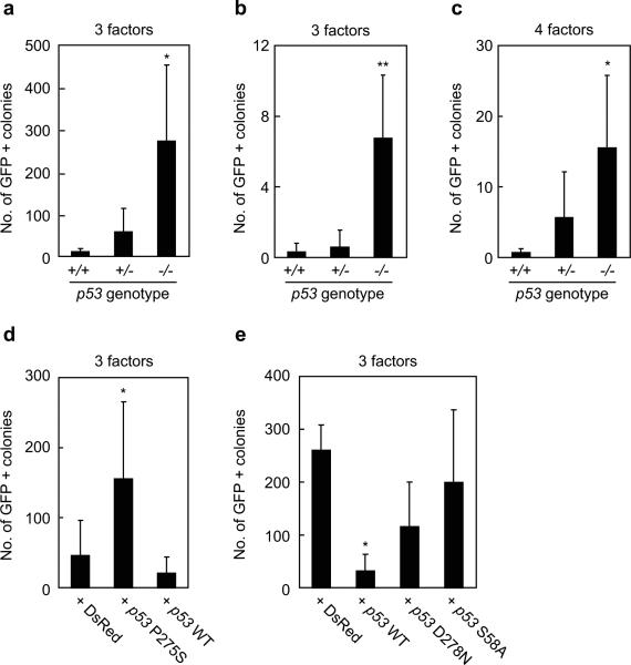
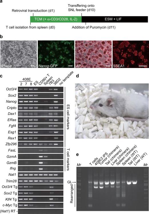
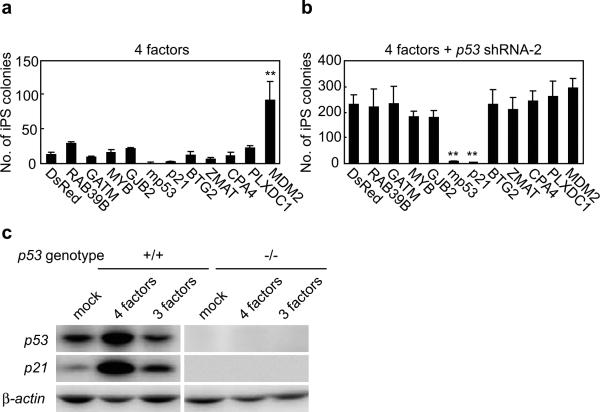
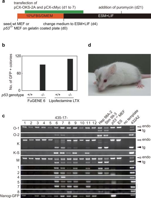
Induced pluripotent stem (iPS) cells can be generated from somatic cells by the introduction of Oct3/4 (also known as Pou5f1), Sox2, Klf4 and c-Myc, in mouse and in human. The efficiency of this process, however, is low. Pluripotency can be induced without c-Myc, but with even lower efficiency. A p53 (also known as TP53 in humans and Trp53 in mice) short-interfering RNA (siRNA) was recently shown to promote human iPS cell generation, but the specificity and mechanisms remain to be determined. Here we report that up to 10% of transduced mouse embryonic fibroblasts lacking p53 became iPS cells, even without the Myc retrovirus. The p53 deletion also promoted the induction of integration-free mouse iPS cells with plasmid transfection. Furthermore, in the p53-null background, iPS cells were generated from terminally differentiated T lymphocytes. The suppression of p53 also increased the efficiency of human iPS cell generation. DNA microarray analyses identified 34 p53-regulated genes that are common in mouse and human fibroblasts. Functional analyses of these genes demonstrate that the p53-p21 pathway serves as a barrier not only in tumorigenicity, but also in iPS cell generation.


Figures

Figure 1
Figure 1. iPS cell generation from p53-null MEF by four or three reprogramming factors
a. iPS cells were generated from Nanog-GFP reporter MEF, which were either p53 wild-type (+/+), heterozygous (+/-), or homozygous (-/-), by the three reprogramming factors (Oct3/4, Sox2, Klf4). After retroviral transduction, 5000 live cells were collected by a flow cytometer. GFP-positive colonies were counted 28 days after the transduction. *, p<0.05 compared to wild-type (n=4). b. iPS cells were generated by the three factors from single sorted cells in wells of 96-well plates. GFP-positive colonies were counted 28 days after the transduction. **, p<0.01 compared to wild-type (n=4). c. iPS cells were generated by the four factors, including c-Myc, from single sorted cells in wells of 96-well plates. GFP-positive colonies were counted 21 days after the transduction. *, p<0.05 compared to wild-type (n=4). d. Retroviruses expressing either the dominant negative p53 mutant (P275S) or wild-type were co-transduced with the three factors into Nanog-GFP, p53 heterozygous MEFs. After retroviral transduction, 5000 cells were collected and GFP-positive colonies were counted 28 days after the transduction. As a control, retrovirus for a red fluorescent protein (DsRed) was transduced. *, p<0.05 compared to DsRed control (n=3). e. Retroviruses expressing either the wild-type or mutant p53 were co-transduced with the three factors into Nanog-GFP, p53-null MEFs. After retroviral transduction, 5000 live cells were collected and GFP-positive colonies were counted 28 days after the transduction. *, p<0.05 compared to DsRed control (n=3).
Figure 2
Figure 2. T-lymphocyte-derived iPS cells
a. Protocol for iPS cell generation from mouse T-lymphocytes. TCM; T cell medium, ESM; ES cell medium. b. A phase contrast image, Nanog-GFP expression, alkaline phosphatase staining, and SSEA1 staining of T cell-derived iPS cells (clone 408E2). Bars indicate 100 μm. c. Expression of marker genes was examined by RT-PCR in T cell-derived iPS cells (clones 408E -2, -7 and -8), RF8 ES cells, T cells, Spleen, Fbx15-selected iPS cells from p53 wild-type MEF (clone 7B3), and Nanog-selected iPS cells from p53 wild-type MEF (clone 38D2) d. A chimera mouse derived from clone 408E2. iPS cells were microinjected into blastocysts from ICR mice. e. The V-(D)-J DNA rearrangement of the Tcrβ gene was confirmed by genomic PCR in iPS cells and a chimeric mouse. GL indicates the germline band.
Figure 3
Figure 3. Effect of c-Myc and p53 suppression on p21
a. Genes regulated by p53 were introduced into HDFs together with the four reprogramming factors. On day 24 post-transduction, numbers of iPS cell colonies were counted. **; p<0.01 compared to DsRed control (n=3). b. Genes regulated by p53 were introduced into HDFs together with the four reprogramming factors and the p53 shRNA. On day 28 post-transduction, numbers of iPS cell colonies were counted. **; p<0.01 compared to DsRed control (n=3). c. Induction of p21 proteins during iPS cell generation. MEFs, either wild-type or p53-null, were tranduced with the four or three reprogramming factors. Three days after the transduction, protein levels of p21 and p53 were determined by western blot analyses.
Figure 4
Figure 4. Effect of p53 suppression on plasmid-mediated mouse iPS cell generation
a. Protocol for mouse iPS cell generation by plasmid transfection. b. Number of GFP-positive colonies. Shown are results of experiment with different transfection reagents. c. Detection of plasmid integration by genomic PCR. d. A chimera mouse derived from integration-free iPS cells. iPS cells were microinjected into blastocysts from ICR mice.

Comment in

References

    1. Takahashi K, Yamanaka S. Induction of pluripotent stem cells from mouse embryonic and adult fibroblast cultures by defined factors. Cell. 2006;126(4):663–676. - PubMed
    1. Maherali N, et al. Directly reprogrammed fibroblasts show global epigenetic remodelling and widespread tissue contribution. Cell Stem Cell. 2007;1(1):55–70. - PubMed
    1. Okita K, Ichisaka T, Yamanaka S. Generation of germ-line competent induced pluripotent stem cells. Nature. 2007;448:313–317. - PubMed
    1. Wernig M, et al. In vitro reprogramming of fibroblasts into a pluripotent ES cell-like state. Nature. 2007;448:318–324. - PubMed
    1. Takahashi K, et al. Induction of pluripotent stem cells from adult human fibroblasts by defined factors. Cell. 2007;131(5):861–872. - PubMed

Publication types

MeSH terms

Substances

Associated data