

doi: 10.1038/nchembio.200.
Epub 2009 Aug 9.
Cell-selective metabolic labeling of proteins
Affiliations
- PMID: 19668194
- PMCID: PMC3176724
- DOI: 10.1038/nchembio.200
Item in Clipboard
Cell-selective metabolic labeling of proteins
Nat Chem Biol.
2009 Oct.
Abstract
Metabolic labeling of proteins with the methionine surrogate azidonorleucine can be targeted exclusively to specified cells through expression of a mutant methionyl-tRNA synthetase (MetRS). In complex cellular mixtures, proteins made in cells that express the mutant synthetase can be tagged with affinity reagents (for detection or enrichment) or fluorescent dyes (for imaging). Proteins made in cells that do not express the mutant synthetase are neither labeled nor detected.
Figures
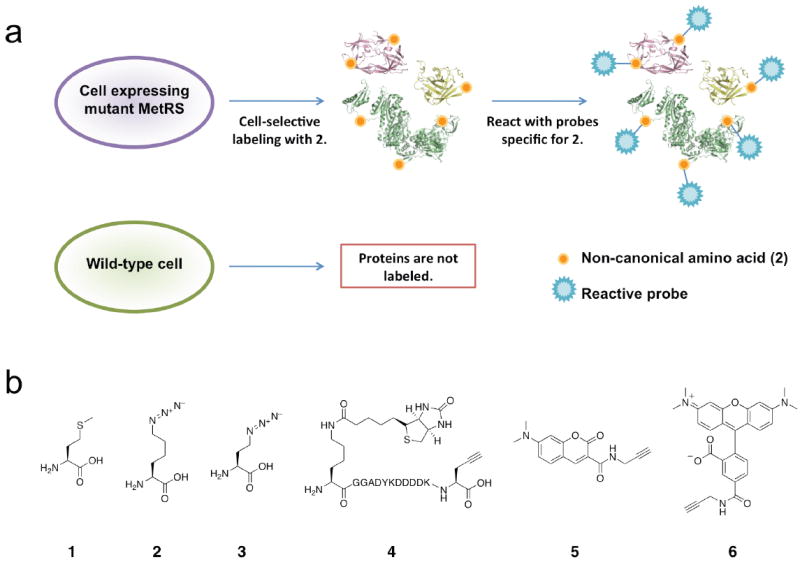
Cell-selective labeling of proteomes with azidonorleucine. (a) Schematic representation of incorporation of azidonorleucine exclusively in cells expressing NLL-MRS. (b) Structures of amino acids and probes used in this study: (1) methionine, (2) azidonorleucine, (3) azidohomoalanine, (4) biotin-FLAG-alkyne, (5) dimethylaminocoumarin-alkyne, (6) TAMRA-alkyne.
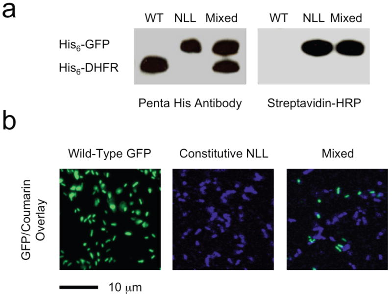
Cell-selective protein labeling in mixed populations. (a) Western blot detection of marker protein expression with Penta-His antibody (left) and with streptavidin-HRP (right). DHFR was made in cells lacking the NLL-MetRS (WT) and GFP in cells expressing the NLL-MetRS (NLL); both proteins contain multiple ATG (Met) codons (7 in DHFR and 8 in GFP). Azidonorleucine was added to all three cultures upon induction of protein synthesis. Samples from the mixed culture contain both proteins (as shown by Penta-His antibody detection), but only GFP is labeled with azidonorleucine and susceptible to labeling with 4. (b) Fluorescence images of cells expressing GFP but not NLL-MetRS (Wild-Type), cells expressing DHFR and NLL-MetRS (NLL), and a mixed culture of the two (Mixed). Azidonorleucine was added to all three cultures upon induction of protein synthesis. Cells from all three cultures were treated with 5, but only cells expressing the NLL-MetRS were labeled. Note that the marker proteins co-expressed with NLL-MetRS are different in the Western blotting and fluorescence imaging experiments.
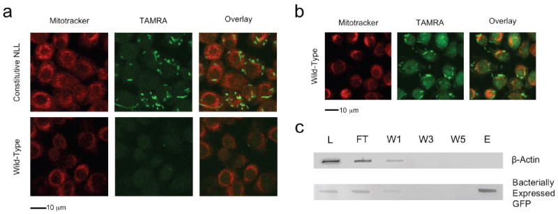
Cell-selective labeling in mixtures of bacterial and mammalian cells. (a) Fluorescence images of mixed cultures containing bacteria attached to or internalized by murine alveolar macrophages. Infection was performed in medium containing azidonorleucine. Bacterial cells constitutively expressing the NLL-MetRS were labeled by TAMRA-alkyne (Constitutive NLL) while cells lacking the NLL-MetRS (Wild type GFP) are visible only in the GFP channel (Supplementary Fig. 4, online). Macrophages were labeled with Mitotracker Deep Red (Invitrogen) and exhibited very low TAMRA background emission. In all cases, conjugation of TAMRA-alkyne was confined to bacterial cells expressing the NLL-MetRS. (b) Fluorescence images of macrophage infection with wild-type bacteria performed in the presence of azidohomoalanine. Protein synthesis by macrophages is indicated by strong TAMRA-alkyne emission from both bacterial cells and macrophages. (c) Macrophages were infected with bacterial cells that express GFP under induction with IPTG and constitutively express the NLL-MetRS. Infection was performed in medium containing IPTG and azidonorleucine to facilitate bacterial synthesis and labeling of GFP. Total cell lysate from the infection was subjected to conjugation with alkyne-functionalized biotin; labeled proteins were enriched with streptavidin avidity. Bacterially expressed GFP and mammalian β-actin were followed by immunoblot. Analysis of the lysate (L), unbound flow-through (FT), washes (W1, W3, W5), and eluent (E) reveal a separation of bacterial and mammalian representative proteins. Bacterially expressed GFP was labeled with azidonorleucine and thus subject to conjugation to biotin and enrichment with streptavidin. Protein originating from macrophages, including β-actin, were not labeled with 2 and therefore not conjugated to alkyne-functionalized biotin.
References
Publication types
MeSH terms
Substances
Grants and funding
LinkOut - more resources
Full Text Sources
Other Literature Sources
