. 2009 Sep;41(9):1032-6.
doi: 10.1038/ng.423. Epub 2009 Aug 9.

Mutations in INPP5E, encoding inositol polyphosphate-5-phosphatase E, link phosphatidyl inositol signaling to the ciliopathies

Affiliations

Mutations in INPP5E, encoding inositol polyphosphate-5-phosphatase E, link phosphatidyl inositol signaling to the ciliopathies

Stephanie L Bielas et al. Nat Genet. 2009 Sep.

Abstract

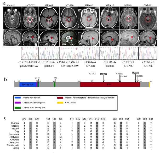
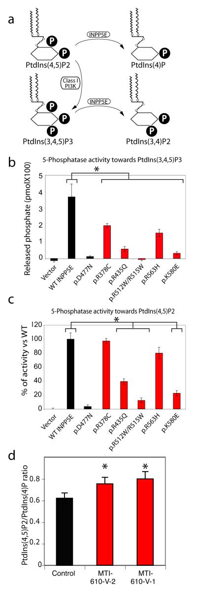
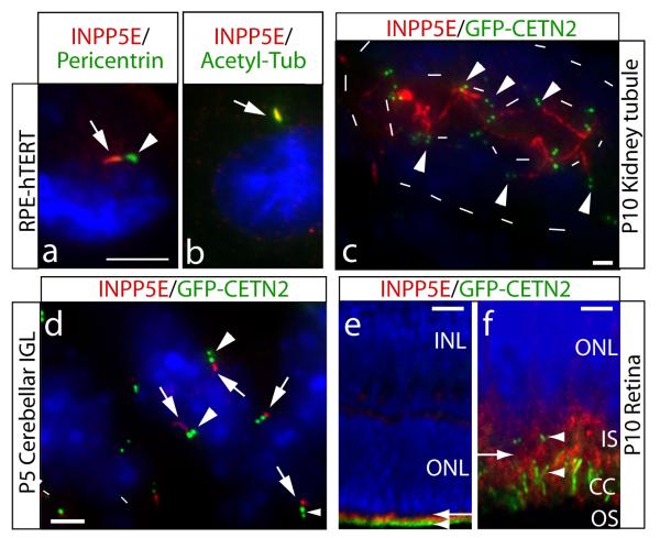
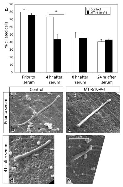
Phosphotidylinositol (PtdIns) signaling is tightly regulated both spatially and temporally by subcellularly localized PtdIns kinases and phosphatases that dynamically alter downstream signaling events. Joubert syndrome is characterized by a specific midbrain-hindbrain malformation ('molar tooth sign'), variably associated retinal dystrophy, nephronophthisis, liver fibrosis and polydactyly and is included in the newly emerging group of 'ciliopathies'. In individuals with Joubert disease genetically linked to JBTS1, we identified mutations in the INPP5E gene, encoding inositol polyphosphate-5-phosphatase E, which hydrolyzes the 5-phosphate of PtdIns(3,4,5)P3 and PtdIns(4,5)P2. Mutations clustered in the phosphatase domain and impaired 5-phosphatase activity, resulting in altered cellular PtdIns ratios. INPP5E localized to cilia in major organs affected by Joubert syndrome, and mutations promoted premature destabilization of cilia in response to stimulation. These data link PtdIns signaling to the primary cilium, a cellular structure that is becoming increasingly recognized for its role in mediating cell signals and neuronal function.


Figures

Figure 1
Figure 1
Missense mutations in the encoded enzymatic domain of INPP5E, (inositol polyphate-5-phosphatase E) in patients linked to the JBTS1 locus. (a) Top: axial brain MRIs showing molar tooth sign (red circle) from one affected from each linked family, representing four different countries of origin. Bottom: midline sagittal MRIs showing horizontally-oriented superior cerebellar peduncle (red arrow), not evident in control. Sequence chromatograms from one affected from each family, showing the nucleotide change (red arrow), with corresponding amino acid substitution listed below. Families MTI-007 and MTI-134 shared a common haplotype at the JBTS1 locus, as did COR-10 and COR-21, and they share common mutations. MTI-007 and MTI-134 have a compound homozygous mutation with a double R512W; R515W. (b) Predicted protein domains of INPP5E indicated by color. Each of the identified missense mutations (arrows) occurs in a basic residue within the catalytic domain and alters charge. (c) Evolutionary conservation of mutated amino acids.
Figure 2
Figure 2
Impaired 5-phosphatase activity and altered ratio of PtdIns(4,5)P2 to PtdIns(4)P associated with JBTS1 INPP5E mutations. (a) Summary of PtdIns metabolism. P = phosphate. A block in INPP5E function is predicted to increase the PtdIns(4,5)P2:PtdIns(4)P ratio. (b-c) More severe reduction in 5-phosphatase activity of mutant INPP5E against PtdIns(3,4,5)P3 than PtdIns(4,5)P2 substrates. Note that activity was largely retained against PtdIns(4,5)2 for some mutations (mutants R435Q, R512W/R515W and K580E are severely defective, whereas R378C and R563H are only slightly diminished). D477N is known phosphatasedead, compared with each of the patient mutations. (N = 3 for each sample) (d) Elevated ratio of PtdIns(4,5)P2 to PtdIns(4)P in patient primary fibroblast lines MTI-610-V-2 and V-1, compared with control fibroblast. * represent p < 0.05 ANOVA two way corrected for multiple comparisons.
Figure 3
Figure 3
Ciliary axonemal localization of INPP5E and enhanced ciliary lability in INPP5E mutant cell line. (a) RPE-hTERT ciliated cells stained for INPP5E and cilia markers. INPP5E (red, arrow) localized adjacent to pericentrin-positive (green, arrowhead) pericentriolar matrix. (b) Colocalization of INPP5E (red, arrow) with ciliary axonemal acetylated tubulin (green). (c-f) show INPP5E (red) staining in GFP-CETN2 transgenic mouse (green pair of centrioles, arrowheads). (c) P10 kidney collecting tubule shows INPP5E-positive red cilia projecting into the renal tubule lumen (inner dashes), limits of tubule indicated by outer dashes. (d) P5 cerebellar internal granule layer with INPP5E (red, arrows) ciliary axoneme adjacent to the centrioles. (e) P10 retina, where GFP-CETN2 labels the basal body and connecting cilium (CC, green, arrowhead). INPP5E exclusively labels photoreceptor cells (red, arrow) (f) High-power view of (e) showing INPP5E staining (red, arrow) in the inner segment (IS), just adjacent to and above basal bodies and CC( arrowheads). Blue = Hoechst. Scale bar 5 um (a-b,d), 25 um (c), 50 um (e), 10 um (f).
Figure 4
Figure 4
(a) Lability of cilia in patient primary fibroblasts with mutant INPP5E. Prior to serum stimulation, serum-starved control and patient fibroblasts displayed comparable percentage of ciliated cells. By 4 hr after serum stimulation, the ciliated cells dropped to 43% in patient fibroblast as compared to 73% in control (* = p < 0.01, N =3, total cells counted >250 for each condition). By 8 hr or 24 hr after stimulation, ciliated cells were again comparable between patient and control samples. (b-e) Scanning electron micrographs of control vs. patient primary fibroblast with mutant INPP5E. Prior to serum stimulation, ciliary axonemal length is comparable, although slightly shorter in patient sample. By 4 hr after stimulation, axonemal length is shorter in patient sample. Scale bar indicated in um.

Similar articles

Cited by

References

    1. Vicinanza M, D’Angelo G, Di Campli A, De Matteis MA. Phosphoinositides as regulators of membrane trafficking in health and disease. Cell Mol Life Sci. 2008 - PMC - PubMed
    1. Valente EM, Brancati F, Dallapiccola B. Genotypes and phenotypes of Joubert syndrome and related disorders. Eur J Med Genet. 2008;51:1–23. - PubMed
    1. Saar K, et al. Homozygosity mapping in families with Joubert syndrome identifies a locus on chromosome 9q34.3 and evidence for genetic heterogeneity. Am J Hum Genet. 1999;65:1666–71. - PMC - PubMed
    1. Valente EM, et al. Distinguishing the four genetic causes of Joubert syndrome-related disorders. Ann Neurol. 2005;57:513–9. - PubMed
    1. Tsujishita Y, Guo S, Stolz LE, York JD, Hurley JH. Specificity determinants in phosphoinositide dephosphorylation: crystal structure of an archetypal inositol polyphosphate 5-phosphatase. Cell. 2001;105:379–89. - PubMed

Publication types

MeSH terms

Substances