. 2009 Sep;41(9):1037-42.
doi: 10.1038/ng.422. Epub 2009 Aug 9.

FOXC1 is required for normal cerebellar development and is a major contributor to chromosome 6p25.3 Dandy-Walker malformation

Affiliations

FOXC1 is required for normal cerebellar development and is a major contributor to chromosome 6p25.3 Dandy-Walker malformation

Kimberly A Aldinger et al. Nat Genet. 2009 Sep.

Abstract

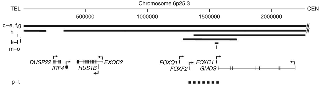
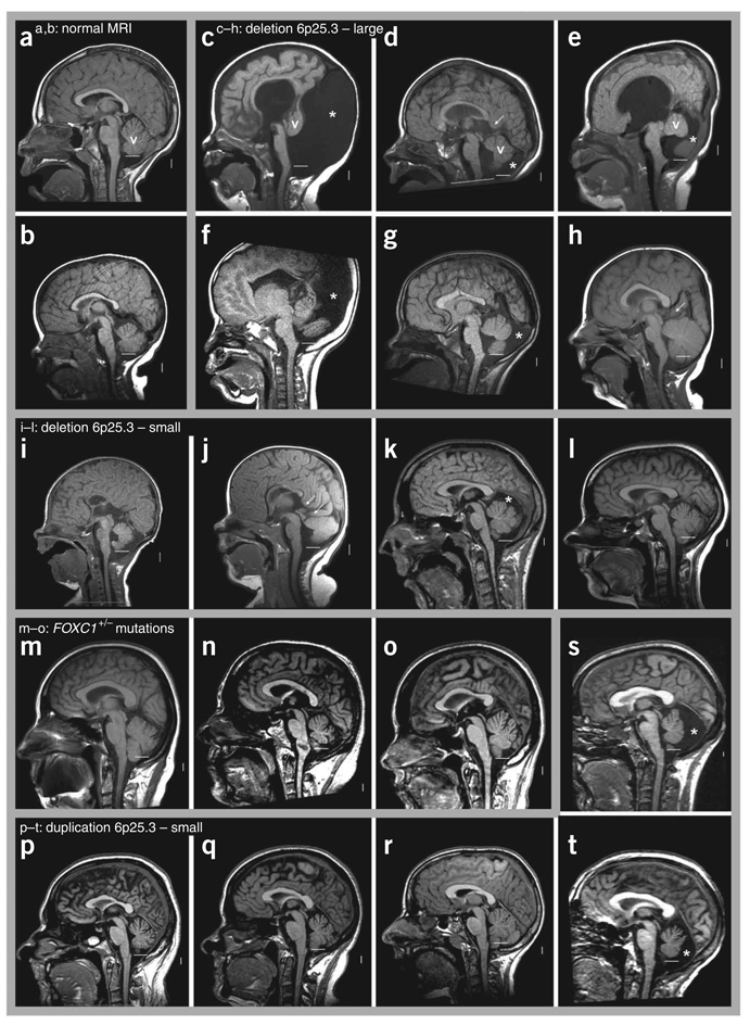
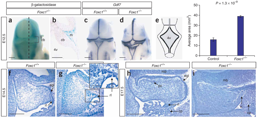
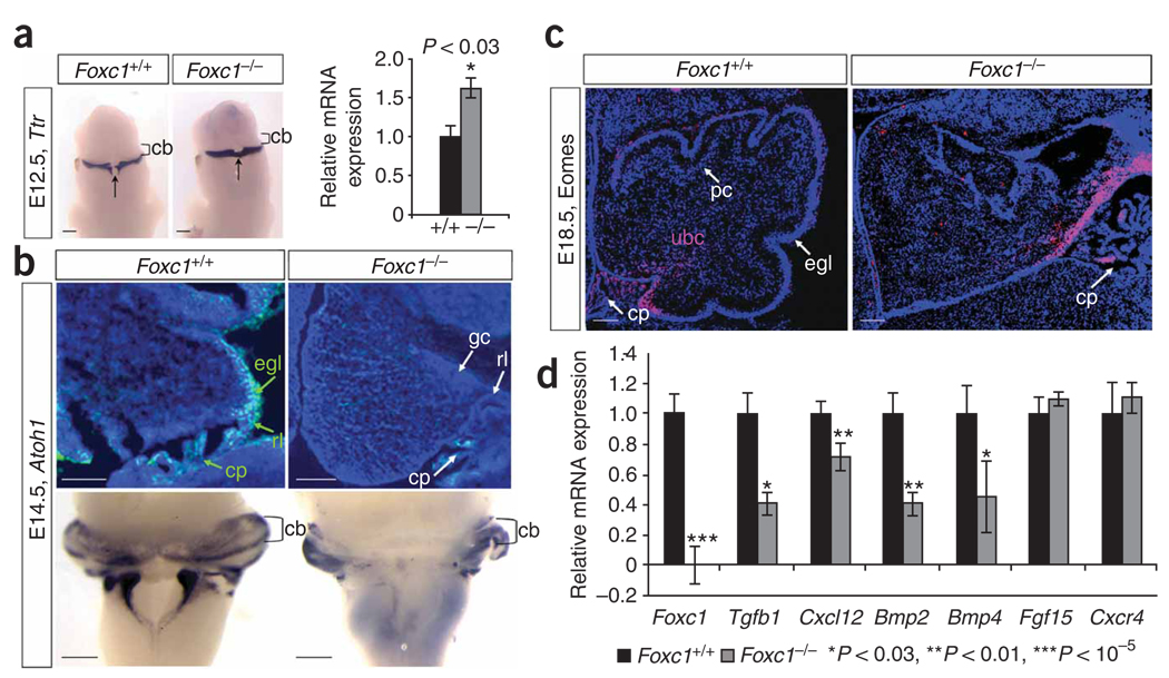
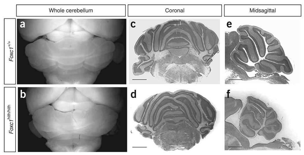
Dandy-Walker malformation (DWM), the most common human cerebellar malformation, has only one characterized associated locus. Here we characterize a second DWM-linked locus on 6p25.3, showing that deletions or duplications encompassing FOXC1 are associated with cerebellar and posterior fossa malformations including cerebellar vermis hypoplasia (CVH), mega-cisterna magna (MCM) and DWM. Foxc1-null mice have embryonic abnormalities of the rhombic lip due to loss of mesenchyme-secreted signaling molecules with subsequent loss of Atoh1 expression in vermis. Foxc1 homozygous hypomorphs have CVH with medial fusion and foliation defects. Human FOXC1 heterozygous mutations are known to affect eye development, causing a spectrum of glaucoma-associated anomalies (Axenfeld-Rieger syndrome, ARS; MIM no. 601631). We report the first brain imaging data from humans with FOXC1 mutations and show that these individuals also have CVH. We conclude that alteration of FOXC1 function alone causes CVH and contributes to MCM and DWM. Our results highlight a previously unrecognized role for mesenchyme-neuroepithelium interactions in the mid-hindbrain during early embryogenesis.


Figures

Figure 1
Figure 1
Physical map of the 6p25.3 DWM locus. Diagram of 6p25.3 drawn to scale shows deletions (solid bars) and duplications (dashed bar) as well as heterozygous mutations of FOXC1 (thin vertical line). Letters to the left of the CNVs refer to the same patients as in Figure 2. RefSeq genes are shown with direction of transcription (arrows). Chromosome position is given in kb.
Figure 2
Figure 2
Brain images of affected individuals within the 6p25.3 cohort. (a–t) T1-weighted midsagittal magnetic resonance images in two control subjects (a,b) and 18 subjects with 6p25.3 CNVs that include FOXC1 or intragenic mutations of FOXC1 (c–t). The midline cerebellar vermis (v) is marked only in the top row of images. The lower limit of the vermis typically extends down to the obex, marked by a horizontal white line in each image. In all but one individual (h), the lower border of the vermis does not reach the white line, indicative of cerebellar vermis hypoplasia (CVH). Asterisks (*) indicate an enlarged posterior fossa. In 5/6 individuals with large deletions of 6p25.3 (c–g), brain images show a small and upwardly rotated vermis, cystic dilatation of the fourth ventricle and an enlarged posterior fossa, meeting criteria for classic Dandy-Walker malformation (DWM). The remaining individual (h) unexpectedly has a normal or mildly enlarged vermis. The vermis appears abnormal in all four individuals with small deletions of 6p25.3 (i–l). The first two (i,j) meet criteria for DWM, although the abnormalities are less severe than in the subject with larger deletions. The next (k) has mild CVH with normal vermis position (not rotated upward), a normal fourth ventricle and mildly enlarged posterior fossa, consistent with mega-cisterna magna (MCM). The last individual in this group (l) has mild CVH only. The three individuals with heterozygous intragenic FOXC1 mutations (m–o) all have mild CVH only. Among the five individuals with 6p25.3 duplication, two sibs (p,t) have obvious MCM, whereas three other sibs—probably distantly related to the first two—have mild CVH only.
Figure 3
Figure 3
Morphological cerebellar phenotype of Foxc1 −/− embryos. (a) Dorsal view of the head from an X-gal–stained Foxc1 +/− (Mf1 +/LacZ) embryo at E12.5 showing that Foxc1 expression excludes the developing brain. (b) Midsagittal section through embryo in a (dashed line) showing expression in the mesenchyme (m) adjacent to the cerebellar anlage (cb). (c) Dorsal view of Gdf7 expression in a wild-type embryo. (d) Dorsal view of Gdf7 expression in a Foxc1 −/− embryo revealing an enlarged fourth ventricle roof plate (4v). (e) Schematic of the dorsal view of an E12.5 embryo depicting the 4v area (shaded gray) measured in Foxc1 −/− and littermates. The 4v was 2.5-fold larger in Foxc1 −/− embryos. (f) Cresyl violet–stained midsagittal section of the developing cerebellar anlage in a wild-type E14.5 embryo. (g) Midsagittal section of the developing cerebellar anlage in a Foxc1 −/− E14.5 embryo showing that the rhombic lip (rl) and external granule cell layer (egl) are abnormal and highly disorganized. (h) Midsagittal section through the cerebellar vermis of a wild-type E17.5 embryo showing distinct egl and Purkinje cell (pc) layers. (i) Midsagittal section through the cerebellar vermis of a Foxc1 −/− E17.5 embryo showing a lack of distinct egl and pc layers, an abnormal rl and an expansion of the choroid plexus (cp). Scale bars, 100 (a,c,d), 50 (b,f–i) or 20 µm (g inset).
Figure 4
Figure 4
Loss of Foxc1 from hindbrain mesenchyme disrupts rhombic lip–derived cerebellar cellular populations. (a) Ttr expression in the differentiating choroid plexus epithelium of E12.5 WT and Foxc1 −/− littermate embryos assayed by in situ hybridization (ISH) shows midline expansion of choroid plexus (arrow) adjacent to the cerebellar anlage (cb) in the mutant. Increased Ttr expression confirmed by qRT-PCR (relative to Gapdh expression) is presented as mean ± s.e.m. *P < 0.03 (b) DAPI-counterstained (blue) parasagittal section shows Atoh1 expression (green) in the cerebellar anlage of E14.5 W Tand Foxc1 −/− littermate embryos. No Atoh1+ cells are observed in the mutant egl. Whole-mount ISH of E14.5 brains confirms that loss of Atoh1 in the mutant is restricted to the developing cerebellar vermis. (c) Eomes expression (pink) in the cerebellar anlage of E18.5 WT and Foxc1 −/− littermate embryos. The Eomes+ unipolar brush cells (ubc) remain in the rl of the mutant, rather than migrating through the developing white matter as in WT. DAPI staining (blue) shows egl and pc in WT cerebellum. (d) Expression of six known Foxc1 downstream targets in eye mesenchyme and endothelial development in the mid-hindbrain of E12.5 WT (black) and Foxc1−/− (gray) littermate embryos, assayed by qRT-PCR. Loss of Foxc1 significantly decreases expression of genes secreted from hindbrain mesenchyme (Tgfb1, Cxcl12, Bmp2 and Bmp4) but not genes expressed in the neural tube (Fgf15 and Cxcr4). Data presented as mean ± s.e.m. represent four independent experiments. *P < 0.03, **P < 0.01, ***P< 0.00001. Scale bars, 100 µm.
Figure 5
Figure 5
Cerebellar morphology of Foxc1 hith/hith mice. (a,b) Dorsal views of mouse brains at postnatal day (P) 30. Mean cerebellar area between Foxc1 hith/hith (5.63 arbitrary units (a.u.) ± 0.31 s.e.m.) and littermates (8.60 a.u. ± 0.03 s.e.m.) reveals significant hypoplasia in these adult mutants (P< 0.01). (c,d) Cresyl violet–stained coronal sections through the cerebellum. (e,f) Cresyl violet–stained midsagittal sections through the vermis of brains of mice at P30. The wild-type cerebellum (a) has a stereotypic foliation pattern, which is disrupted in the homozygous mutants. Scale bars, 200 µm.

References

    1. Millen KJ, Gleeson JG. Cerebellar development and disease. Curr. Opin. Neurobiol. 2008;18:12–19. - PMC - PubMed
    1. Grinberg I, et al. Heterozygous deletion of the linked genes ZIC1 and ZIC4 is involved in Dandy-Walker malformation. Nat. Genet. 2004;36:1053–1055. - PubMed
    1. Parisi MA, Dobyns WB. Human malformations of the midbrain and hindbrain: review and proposed classification scheme. Mol. Genet. Metab. 2003;80:36–53. - PubMed
    1. Patel S, Barkovich AJ. Analysis and classification of cerebellar malformations. AJNR Am. J. Neuroradiol. 2002;23:1074–1087. - PMC - PubMed
    1. DeScipio C, et al. Subtelomeric deletions of chromosome 6p: molecular and cytogenetic characterization of three new cases with phenotypic overlap with RitscherSchinzel (3C) syndrome. Am. J. Med. Genet. A. 2005;134:3–11. - PubMed

Publication types

MeSH terms

Substances