. 2009 Oct 22;28(42):3735-45.
doi: 10.1038/onc.2009.232. Epub 2009 Aug 10.

The prolyl isomerase Pin1 regulates the NF-kappaB signaling pathway and interleukin-8 expression in glioblastoma

Affiliations

The prolyl isomerase Pin1 regulates the NF-kappaB signaling pathway and interleukin-8 expression in glioblastoma

G P Atkinson et al. Oncogene. .

Abstract

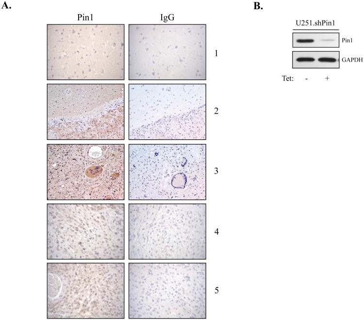
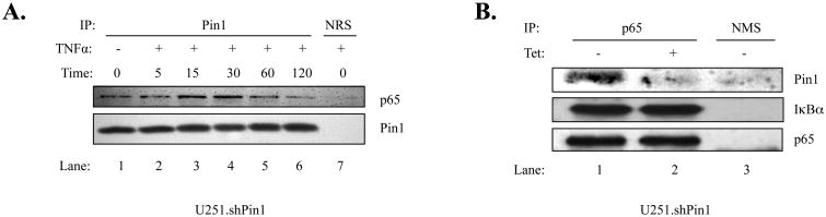
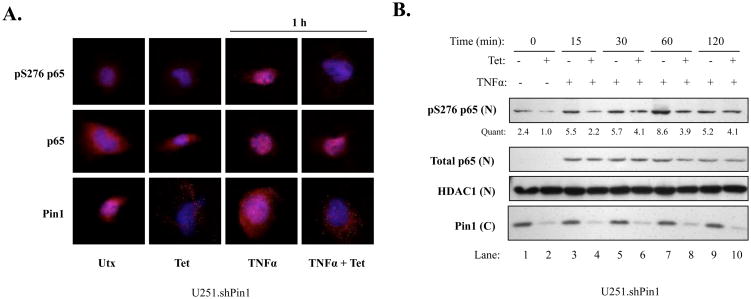
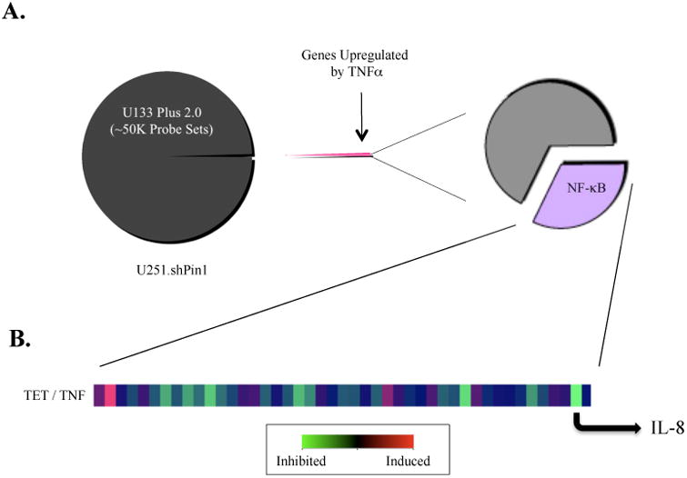
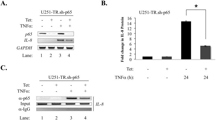
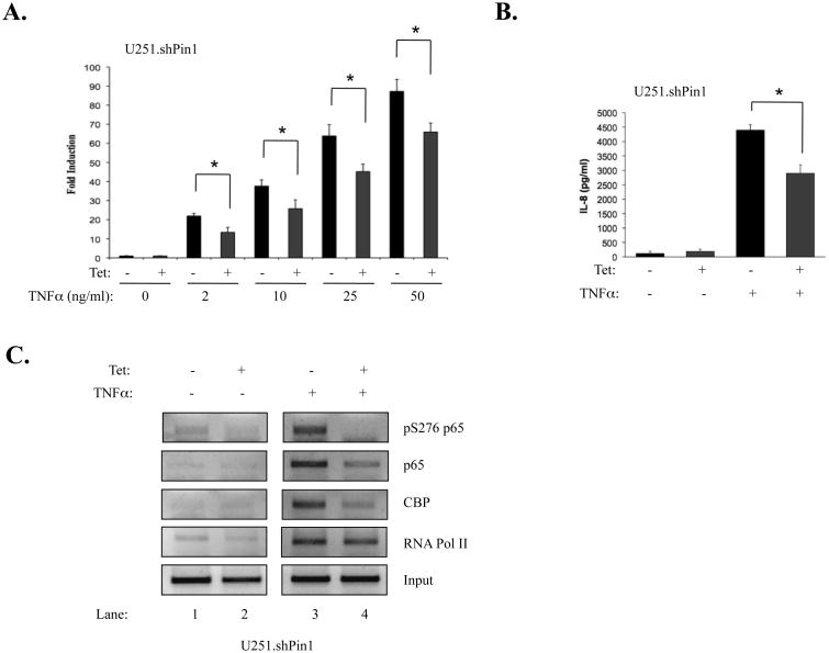
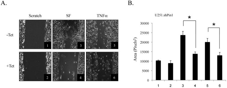
The brain tumor glioblastoma (GBM) remains one of the most aggressive and devastating tumors despite decades of effort to find more effective treatments. A hallmark of GBM is the constitutive activation of the nuclear factor kappa-light-chain-enhancer of activated B cells (NF-kappaB) signaling pathway, which regulates cell proliferation, inflammation, migration and apoptosis. The prolyl isomerase, Pin1, has been found to bind directly to the NF-kappaB protein, p65, and cause increases in NF-kappaB promoter activity in a breast cancer model. We now present evidence that this interaction occurs in GBM and that it has important consequences on NF-kappaB signaling. We demonstrate that Pin1 levels are enhanced in primary GBM tissues compared with controls, and that this difference in Pin1 expression affects the migratory capacity of GBM-derived cells. Pin1 knockdown decreases the amount of activated, phosphorylated p65 in the nucleus, resulting in inhibition of the transcriptional program of the IL-8 gene. Through the use of microarray, we also observed changes in the expression levels of other NF-kappaB regulated genes due to Pin1 knockdown. Taken together, these data suggest that Pin1 is an important regulator of NF-kappaB in GBM, and support the notion of using Pin1 as a therapeutic target in the future.


Figures

Figure 1
Figure 1. Pin1 Expression in Primary Human Tissues and Cell Lines
(A) Immunohistochemistry was performed on control and GBM paraffin-embedded brain tissue. Each section was stained with anti-Pin1 antibody and an IgG control antibody. A total of ten tumor samples and eight control brain samples were analyzed, and representative images from one control sample (1) and four GBM samples (2-5) are shown. (B) U251.shPin1 cells were grown in the absence or presence of Tet (5 μg/ml) for 96 h. Total cell lysates were assayed by immunoblot with anti-Pin1 and anti-GAPDH antibodies. Representative of three experiments.
Figure 2
Figure 2. Pin1 Associates with p65 in U251-MG Cells
(A) U251 shPin1 cells were grown in 150 mm dishes. When cell density reached approximately 80%, the cells were serum-starved for 4 h, treated with TNFα (10 ng/ml) for the indicated times (0-120 min), and lysed in RIPA buffer. Lysate from each sample was then subjected to immunoprecipitation (IP) using antibodies specific for Pin1 or normal rabbit serum (NRS). IP samples were probed with antibodies to p65 to demonstrate a specific interaction between p65 and Pin1. (B) U251.shPin1 cells were grown in 150 mm dishes in the absence or presence of Tet for 72 h. Cells were lysed in RIPA buffer and anti-p65 was used in the subsequent IP along with a normal mouse serum (NMS) control. Immunoblotting was performed using antibodies against Pin1, IκBα and p65. Results shown are representative of three experiments.
Figure 3
Figure 3. Pin1 Knockdown Inhibits S276 Phosphorylation of p65
(A) U251 shPin1 cells were grown in the absence or presence of Tet for 72 h. 1 × 10⁴ cells were plated in 8-well chamber slides and allowed to recover overnight. Cells were then serum starved, stimulated with 10 ng/ml of TNFα for 1 h, and assayed by immunofluorescence with the indicated antibodies. Dapi was used to stain nuclei (blue) and the indicated antibodies were used to analyze protein levels and localization (red). Multiple images were taken from each chamber; representative images for each condition from three experiments are shown. (B) 2.7 × 10⁵ U251.shPin1 cells were plated in 100 mm dishes and grown in the absence or presence of Tet. After 72 h, media was changed to serum-free and cells were serum-starved overnight. The next day, cells were stimulated with 50 ng/ml of TNFα for the given timepoints and washed once with PBS. Nuclear (N) and cytoplasmic (C) fractions were separated using the Pierce NE-PER Kit and assayed via immunoblotting with the indicated antibodies. Quantification (Quant) of nuclear pS276 p65 was calculated by dividing the densitometric value of each lane by the corresponding nuclear HDAC1 value. Results are representative of three experiments.
Figure 4
Figure 4. Microarray Data Overview
(A) Three conditions were compared: cells with no treatment (UTX), cells treated with TNFα only (TNF), and cells treated with both TNFα and Tet (TET). Approximately 50,000 genes were filtered, allowing only genes that were significantly upregulated (four-fold increase) after a 4 h treatment with TNFα to pass through. This daughter set was cross-referenced with a comprehensive database of NF-κB regulated genes from NF-κB.org, and these NF-κB regulated genes were then analyzed for changes in gene expression due to Pin1 knockdown. (B) A heat-map depicting the ratio TET / TNF shows changes in expression levels of NF-κB regulated genes. IL-8 was selected as a strong candidate for further evaluation due to its inhibition upon depletion of Pin1.
Figure 5
Figure 5. p65 Knockdown Inhibits IL-8 Expression and Decreases p65 Recruitment to the IL-8 Promoter
(A) U251-TR.sh-p65 cells were grown in the absence or presence of Tet for 48 h, serum-starved, and then stimulated with TNFα (10 ng/ml) for 4 h. Total RNA was isolated, first-strand cDNA synthesis was performed, and RT-PCR was performed on the samples using primers to specifically detect p65, IL-8 and GAPDH. (B) U251-TR.sh-p65 cells were grown in the absence or presence of Tet for 48 h, serum-starved, and the stimulated with TNFα (10 ng/ml) for 24 h. Supernatant was collected and analyzed by ELISA for IL-8 protein expression (*p<0.05). (C) U251-TR.sh-p65 cells were grown in the absence or presence of Tet for 48 h, serum-starved, and then stimulated with TNFα (10 ng/ml) for 15 min. The samples were then subjected to ChIP assay (see Materials and Methods). Immunoprecipitations were performed with anti-p65 or an IgG control. All results are representative of three experiments.
Figure 6
Figure 6. Pin1 Knockdown Inhibits IL-8 Gene Expression and Decreases p65 Recruitment at the IL-8 Promoter
(A) Two-step quantitative RT-PCR was performed using SYBR Green. One μg of RNA was reverse transcribed to cDNA as described. Mastermix, primers, and cDNA were combined and run for 50 cycles in an ABI 7500. Ct values for IL-8 qPCR reactions were derived and divided by Ct values for GAPDH. Results are representative of three experiments (*p<0.05). (B) Pin1 knockdown inhibits IL-8 protein expression. Cells were plated at 2.0 × 10⁴ in 12-well plates, grown in the absence or presence of Tet, and stimulated for 24 h with TNFα. Supernatants were analyzed for IL-8 protein expression by ELISA. Results are representative of three experiments (*p<0.05). (C) 3.0 × 10⁶ U251.shPin1 cells were grown in the absence or presence of Tet for 72 h, plated in 150 mm dishes, serum-starved, and stimulated with TNFα. Cells were assayed by ChIP (see Materials and Methods). Immunoprecipitations were performed with antibodies to the given proteins and primers specific to the IL-8 promoter. A PCR reaction using the human IL-8 promoter primers on non-immunoprecipitated DNA (Input) was included as a control for total DNA levels.
Figure 7
Figure 7. Pin1 Knockdown Decreases the Migration of U251-MG Cells
(A) Cells were grown in the absence or presence of Tet (5 μg/ml) for 72 h and plated in 6-well plates. Wells were then scratched with a sterile p200 pipette tip, washed with PBS, and changed to serum-free media (SF) for 48 h. Conditions 5 and 6 were treated with TNFα for 36 h. Conditions were performed in triplicate and three images were taken of each replicate. Representative images from each condition are shown. (B) Images from the scratch assay were quantified using ImageJ software. Results are representative of three experiments (*p<0.05).

References

    1. Aggarwal BB. Nuclear factor-kappaB: the enemy within. Cancer Cell. 2004;6:203–8. - PubMed
    1. Ayala G, Wang D, Wulf G, Frolov A, Li R, Sowadski J, et al. The prolyl isomerase Pin1 is a novel prognostic marker in human prostate cancer. Cancer Res. 2003;63:6244–51. - PubMed
    1. Baker BJ, Qin H, Benveniste EN. Molecular basis of oncostatin M-induced SOCS-3 expression in astrocytes. Glia. 2008;56:1250–62. - PMC - PubMed
    1. Bao L, Kimzey A, Sauter G, Sowadski JM, Lu KP, Wang DG. Prevalent overexpression of prolyl isomerase Pin1 in human cancers. Am J Pathol. 2004;164:1727–37. - PMC - PubMed
    1. Bharti AC, Aggarwal BB. Nuclear factor-kappa B and cancer: its role in prevention and therapy. Biochem Pharmacol. 2002;64:883–8. - PubMed

Publication types

MeSH terms