

Ocular myositis: diagnostic assessment, differential diagnoses, and therapy of a rare muscle disease - five new cases and review
- PMID: 19668464
- PMCID: PMC2699981
Ocular myositis: diagnostic assessment, differential diagnoses, and therapy of a rare muscle disease - five new cases and review
Abstract
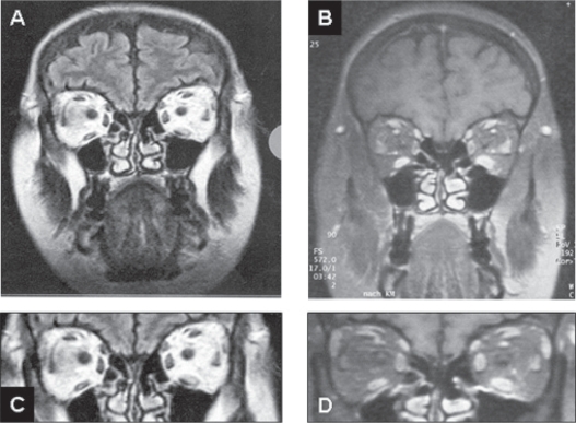
Ocular myositis represents a subgroup within the idiopathic orbital inflammatory syndrome, formerly termed orbital pseudotumor. Ocular myositis describes a rare inflammatory disorder of single or multiple extraocular eye muscles. Unilateral or sequential bilateral subacute painful diplopia is the leading symptom of eye muscle myositis. There are at least two major forms, a limited oligosymptomatic ocular myositis (LOOM) with additional conjunctival injections only, and a severe exophthalmic ocular myositis (SEOM) with additional ptosis, chemosis, and proptosis. Eye muscle myositis is an idiopathic inflammation of the extraocular muscles in the absence of thyroid disease, ocular myasthenia gravis, and other systemic, particularly autoimmune mediated diseases, resembling CD4(+) T cell-mediated dermatomyositis. Contrast-enhanced orbital magnetic resonance imaging most sensitively discloses swelling, signal hyperintensity, and enhancement of isolated eye muscles. Typically, corticosteroid treatment results in prompt improvement and remission within days to weeks in most patients. Compiled data of five patients and a review of the clinical pattern, diagnostic procedures, differential diagnoses, and current treatment options are given.
Keywords: enlarged extraocular muscles; idiopathic orbital inflammation; ocular myositis; painful diplopia.
Figures
References
-
- Bau V, Zierz S. Update on chronic progressive external ophthalmoplegia. Strabismus. 2005;13:133–42. - PubMed
-
- Beeson D, Hantai D, Lochmuller H, et al. 126th International Workshop: congenital myasthenic syndromes, 24–26 September 2004, Naarden, the Netherlands. Neuromuscul Disord. 2005;15:498–512. - PubMed
-
- Berkhoff M, Sturzenegger M, Schroth G, et al. Ocular myositis. Nervenarzt. 1997;68:792–800. - PubMed
-
- Birch-Hirschfeld A. [Handbuch der Gesamten Augenheilkunde] Vol. 9. Berlin: Julius Springer; 1930. p. 251.
-
- Brais B. Oculopharyngeal muscular dystrophy: a late-onset polyalanine disease. Cytogenet Genome Res. 2003;100:252–60. - PubMed
LinkOut - more resources
Full Text Sources
Research Materials
