. 2007 Mar;1(1):43-53.

Apical localization of glutamate in GLAST-1, glutamine synthetase positive ciliary body nonpigmented epithelial cells

Affiliations
Free PMC article

Apical localization of glutamate in GLAST-1, glutamine synthetase positive ciliary body nonpigmented epithelial cells

Marlyn P Langford et al. Clin Ophthalmol. 2007 Mar.
Free PMC article

Abstract

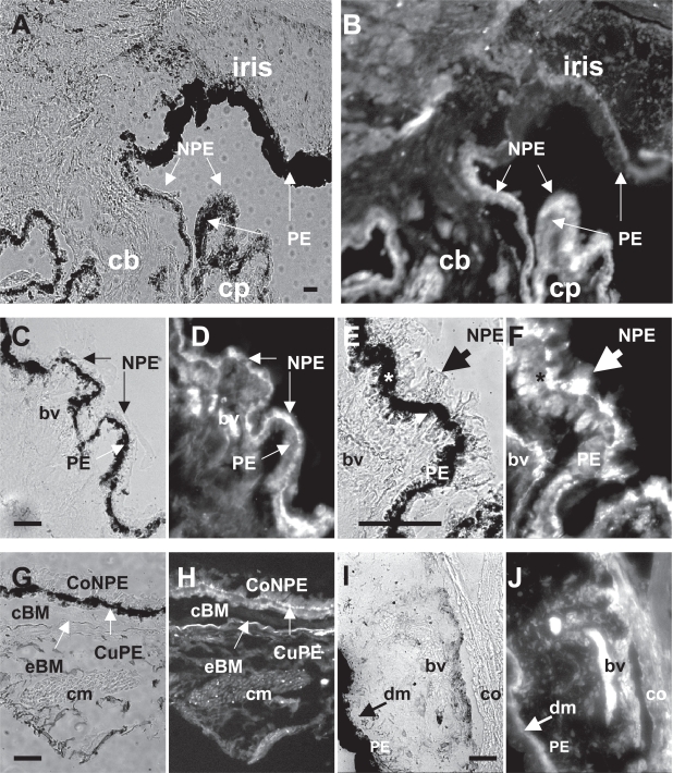
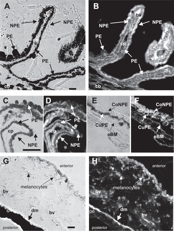
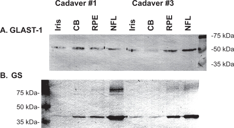
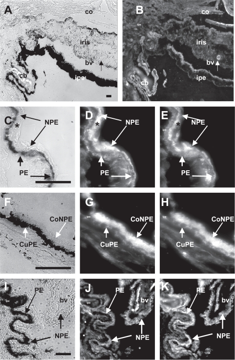
The distribution of glutamate (Glu), the Glu transporter GLAST-1, and glutamine synthetase (GS) in human and monkey anterior uveal tissue, as well as serum (S) to aqueous humor (AH) Glu and glutamine (Gln) gradients were investigated. Cross-linked Glu (xGlu), GLAST-1, and GS were detected using the immunofluorescent antibody technique. S/AH Glu, Gln, and alanine (Ala) concentrations were quantified by high performance liquid chromatography. xGlu immunoreactivity was detected in melanocytes, posterior pigmented epithelial/dilator muscle cells, vascular endothelial cells, and lymphocytes of the iris, as well as the pigmented (PE) and nonpigmented epithelial (NPE) cells and muscle cells of ciliary body. xGlu immunoreactivity was highly concentrated at the apices of GLAST-1, GS positive ciliary body NPE cells, and in GLAST-1 positive iris melanocytes and iris dilator muscle cells. AH Glu concentrations were lower (p < 0.001), while Gln was higher in monkey (p = 0.01) and human cataractous (p = 0.15) AH than serum. The results indicate that Glu is concentrated within GLAST-1, GS positive NPE cells and are consistent with the suggestion that Glu and Gln concentrations in AH may be due in part to GLAST-1 and GS activity in iris and ciliary body epithelial cells.

Keywords: ciliary body; eye; glutamate; glutamine synthetase; nonpigmented epithelial cells; uvea.


Figures

Figure 1
Figure 1
Distribution of xGlu immunoreactivity in anterior uveal tissues from human cadaver eyes. (A) Light photomicrograph of the inis/ciliary body (cb) of the #3 human eye (Table 1) and corresponding (B) IFA photomicrograph showing xGlu immunoreactivity in epithelial cells that line the anterior and posterior iris and ciliary body. Note the intense immunofluorescence staining of the ciliary process (cp) epithelial cells. (C) High magnification light photomicrograph of human ciliary body pars plicata tissue from the #2 donor eye and (D) corresponding IFA photomicrograph showing the intense xGlu immunoreactivity signal in NPE cells. (E) Higher magnification light photomicrograph of human ciliary body tissue from the #1 donor eye and (F) the corresponding IFA photomicrograph showing intense xGlu immunoreactivity in PE and NPE cells (*). Note the highly intense immunofluorescent signal at the apical surface of the NPE cells adjacent to the apical surface of the PE cells (large arrow). (G) Light photomicrograph of human pars plana tissue from the #3 donor eye and corresponding (H) IFA photomicrograph showing xGlu immunoreactivity in the columnar NPE (CoNPE) and cuboidal PE (CuPE) cells of the pars plana. Also, note the high concentration of xGlu immunoreactivity at the apical surface of the CoNPE cells and in ciliary smooth muscle (cm) cells of the #3 donor eye. (I) Light photomicrograph of #2 donor iris and the corresponding (J) IFA photomicrograph showing xGlu immunoreactivity in the cornea (co) cells, iris pigment epithelium and dilator muscle (dm) cell layer, thick walled blood vessels (bv), melanocytes of the vascular layer, and pigment cells on the anterior border of the iris surface. (Bar = 50 μm). Abbreviations: bv, blood vessel; cb, ciliary body; cm, ciliary muscle; CoNPE, columnar NPE; cp, ciliary process; CuPE, cuboidal PE; cBM, cuticle layer of Bruch’s membrane; dm, dilator muscle; eBM, elastic layer of Bruch’s membrane; Gln, glutamine; IFA, immunofluorescent-antibody; NPE, nonpigmented epithelial; PE, pigmented epithelial; xGlu, cross-linked glutamate.
Figure 2
Figure 2
Distribution of xGlu immunoreactivity in monkey ciliary body (cb) and iris tissue. (A) Light photomicrograph and (B) IFA showing xGlu immunoreactivity in the PE and NPE cells of #1 monkey pars plicata ciliary processes (cp). Note the higher concentration of xGlu immunoreactivity at the apical surface of some of the NPE cells (*). (C) Light photomicrograph of a high power magnification of a section of #2 monkey ciliary body tissue and (D) the corresponding IFA section showing xGlu immunoreactivity in PE and NPE cells with high concentration of xGlu immunoreactivity at the apices of some NPE cells (*). (E) Light photomicrograph of pars plana tissue and (F) the corresponding IFA showing a similar distribution of concentrated xGlu immunoreactivity in cuPE and coNPE cells (anterior to the elastic Brock’s membrane; eBM) as in para plana tissue. (G) Light photomicrograph of iris tissue and (H) corresponding IFA showing xGlu immunoreactivity in the anterior iris cells, iris melanocytes, blood vessel (bv) cells, and pigment containing epithelial/dilator muscle (dm) cells. (Bar = 50 mm). Abbreviations: bv, blood vessel; cb, ciliary body; cm, ciliary muscle; CoNPE, columnar NPE; cp, ciliary process; CuPE, cuboidal PE; cBM, cuticle layer of Bruch’s membrane; dm, dilator muscle; eBM, elastic layer of Bruch’s membrane; Gln, glutamine; IFA, immunofluorescent-antibody; NPE, nonpigmented epithelial; PE, pigmented epithelial; xGlu, cross-linked glutamate.
Figure 3
Figure 3
Western blot analysis for (A) GLAST-1 and (B) GS protein expression in iris, ciliary body (CB), retinal pigmented epithelium (RPE) and nerve fiber layer (NFL) tissues from 2 human cadaver eyes (5 μg protein/lane). Abbreviations: GLAST-1, a glutamate transporter; GS, glutamine synthetase.
Figure 4
Figure 4
Distribution of GLAST-1 and GS with xGlu immunoreactivity in human anterior uveal tissue. (A) Light microscopy of a human iris and ciliary body (cb) tissue section from the #2 cadaver eye. (B). Fluorescent microscopy of the same tissue section showing intense GLAST-1 immunoreactivity in cornea endothelium (co), thick walled iris blood vessel (bv), iris pigment epithelium/dilator muscle cells (ipe), and PE and NPE cells that line the ciliary body (cb). (C) High magnification of ciliary body epithelia. (D) High power of the same section showing the distribution of immunoreactive GLAST-1 to the basal surface of PE and the basal and apical surfaces of NPE cells. (E) Corresponding high power magnification of the same section showing the distribution of xGlu immunoreactivity in PE and NPE cells with concentrated xGlu immunoreactivity localized to the apical side of NPE cells juxtaposed to the melanosome at the apical side of the PE cells. Note that GLAST-1 was detected on the basal PE and apical NPE surfaces and was associated with concentrated xGlu immunoreactivity (*) in NPE cells. (F) High power light microscopy of human pars plana ciliary body tissue. (G) Corresponding fluorescent microscopy showing cuboidal PE (cuPE) and columnar NPE (coNPE) cells positive for GLAST-1. (H) Distribution of xGlu immunoreactivity in the same pars plana tissue section. (I) High power light microscopy of #3 donor ciliary body tissue with (J) corresponding fluorescent microscopy showing blood vessels, PE and NPE cells positive for GS immunoreactivity. (K) Distribution of xGlu immunoreactivity in the same section. Note that xGlu positive NPE cells were positive for GS (Bar = 50 μm). Abbreviations: GLAST-1, a glutamate transporter; GS, glutamate synthetase; IFA, immunofluorescent-antibody; NPE, nonpigmented epithelial; PE, pigmented epithelial; xGlu, cross-linked glutamate.

References

    1. Arriza JL. Functional comparisons of three glutamate transporter subtypes cloned from motor cortex. J Neurosci. 1994;14:5559–69. - PMC - PubMed
    1. Arriza JL, Eliasof S, Kavanaugh MP, et al. Excitatory amino acid transporter 5, a retinal glutamate transporter coupled to a chloride conductance. Proc Natl Acad Sci U S A. 1997;94:4155–60. - PMC - PubMed
    1. Barnett NL, Pow DV, Robinson SR. Inhibition of Müller cell glutamine synthetase rapidly impairs the retinal response to light. Glia. 2000;30:64–73. - PubMed
    1. Bridges CC, Kekuda R, Wang H, et al. Structure, function, and regulation of human cystine/glutamate transporter in retinal pigment epithelial cells. Invest Ophthalmol Vis Sci. 2001;42:47–52. - PubMed
    1. Choi DW, Maulucci-Gedde M, Kriegstein AR. Glutamate neurotoxicity in cortical cell culture. J Neurosci. 1987;7:357–68. - PMC - PubMed

LinkOut - more resources