

Clinical approach to optic neuropathies
- PMID: 19668477
- PMCID: PMC2701125
Clinical approach to optic neuropathies
Abstract
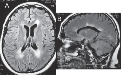
Optic neuropathy is a frequent cause of vision loss encountered by ophthalmologist. The diagnosis is made on clinical grounds. The history often points to the possible etiology of the optic neuropathy. A rapid onset is typical of demyelinating, inflammatory, ischemic and traumatic causes. A gradual course points to compressive, toxic/nutritional and hereditary causes. The classic clinical signs of optic neuropathy are visual field defect, dyschromatopsia, and abnormal papillary response. There are ancillary investigations that can support the diagnosis of optic neuropathy. Visual field testing by either manual kinetic or automated static perimetry is critical in the diagnosis. Neuro-imaging of the brain and orbit is essential in many optic neuropathies including demyelinating and compressive. Newer technologies in the evaluation of optic neuropathies include multifocal visual evoked potentials and optic coherence tomography.
Keywords: Leber’s optic neuropathy; arteritic anterior ischemic optic neuropathy (AION); dominant optic atrophy; multiple sclerosis; non-arteritic anterior ischemic optic neuropathy (NAION); optic neuritis; optic neuropathy; optical coherence tomography; radiation optic neuropathy; recessive optic atrophy; traumatic optic neuropathy.
Figures
References
-
- Ajax ET, Kardon R. Late-onset Leber’s hereditary optic neuropathy. J Neuroophthalmol. 1998;18:30–1. - PubMed
-
- Ajlouni K, Jarrah N, et al. Wolfram syndrome: identification of a phenotypic and genotypic variant from Jordan. Am J Med Genet. 2002;115:61–5. - PubMed
-
- Arnold AC. Evolving management of optic neuritis and multiple sclerosis. Am J Ophthalmol. 2005;139:1101–8. - PubMed
-
- Arnold AC, Hepler RS. Natural history of nonarteritic anterior ischemic optic neuropathy. J Neuroophthalmol. 1994;14:66–9. - PubMed
-
- Asproudis IC, Nikas AN, et al. Paraneoplastic optic neuropathy in a patient with a non-small cell lung carcinoma: a case report. Eur J Ophthalmol. 2005;15:420–3. - PubMed
LinkOut - more resources
Full Text Sources
Other Literature Sources
Medical
