Recent advances in corneal regeneration and possible application of embryonic stem cell-derived corneal epithelial cells
- PMID: 19668514
- PMCID: PMC2704521
Recent advances in corneal regeneration and possible application of embryonic stem cell-derived corneal epithelial cells
Abstract
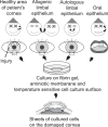
The depletion of limbal stem cells due to various diseases leads to corneal opacification and visual loss. The unequivocal identification and isolation of limbal stem cells may be a considerable advantage because long-term, functional recovery of corneal epithelium is linked to graft constructs that retain viable stem cell populations. As specific markers of limbal stem cells, the ATP-binding cassette, sub-family G, member2 (ABCG2), a member of the multiple drug-resistance (MDR) family of membrane transporters which leads to a side population phenotype, and transcription factor p63 were proposed recently. Conventional corneal transplantation is not applicable for patients with limbal stem cells deficiency, because the conventional allograft lacks limbal stem cells. The introduction of limbal epithelial cell transplantation was a major advance in the therapeutic techniques for reconstruction of the corneal surface. Limbal epithelial cell transplantation is clinically conducted when cultured allografts as well as autografts are available; however, allografts have a risk of immunologic rejection and autografts are hardly available for patients with bilateral ocular surface disorders. Embryonic stem (ES) cells are characterized by their capacity to proliferate indefinitely and to differentiate into any cell type. We induced corneal epithelial cells from ES cells by culturing them on type IV collagen or alternatively, by introduction of the pax6 gene into ES cells. Recent advances in our study supports the possibility of their clinical use as a cell source for reconstruction of the damaged corneal surface. This review summarizes the recent advances in corneal regeneration therapies and the possible application of ES cell-derived corneal epithelial cells.
Keywords: corneal epithelial cell; embryonic stem cell; limbal stem cell; transplantation.
Figures


Similar articles
-
Identification and characterization of limbal stem cells.Exp Eye Res. 2005 Sep;81(3):247-64. doi: 10.1016/j.exer.2005.02.016. Exp Eye Res. 2005. PMID: 16051216 Review.
-
Influence of feeder layer on the expression of stem cell markers in cultured limbal corneal epithelial cells.Indian J Med Res. 2008 Nov;128(5):616-22. Indian J Med Res. 2008. PMID: 19179682
-
Characterization, isolation, expansion and clinical therapy of human corneal epithelial stem/progenitor cells.J Stem Cells. 2014;9(2):79-91. J Stem Cells. 2014. PMID: 25158157 Review.
-
Ex vivo expansion of corneal limbal epithelial/stem cells for corneal surface reconstruction.Eur J Ophthalmol. 2003 Jul;13(6):515-24. doi: 10.1177/112067210301300602. Eur J Ophthalmol. 2003. PMID: 12948308 Review.
-
Reconstruction of damaged corneal epithelium using Venus-labeled limbal epithelial stem cells and tracking of surviving donor cells.Exp Eye Res. 2013 Oct;115:246-54. doi: 10.1016/j.exer.2013.07.024. Epub 2013 Aug 8. Exp Eye Res. 2013. PMID: 23933569
Cited by
-
Recovering vision in corneal epithelial stem cell deficient eyes.Cont Lens Anterior Eye. 2019 Aug;42(4):350-358. doi: 10.1016/j.clae.2019.04.006. Epub 2019 Apr 29. Cont Lens Anterior Eye. 2019. PMID: 31047800 Free PMC article. Review.
-
One year of Clinical Ophthalmology.Clin Ophthalmol. 2007 Dec;1(4):353. Clin Ophthalmol. 2007. PMID: 19668511 Free PMC article. No abstract available.
-
Hyaluronan supports the limbal stem cell phenotype during ex vivo culture.Stem Cell Res Ther. 2022 Jul 30;13(1):384. doi: 10.1186/s13287-022-03084-8. Stem Cell Res Ther. 2022. PMID: 35907870 Free PMC article.
References
-
- Akle CA, Adinolfi M, Welsh KI, et al. Immunogenicity of human amniotic epithelial cells after transplantation into volunteers. Lancet. 1981;2:1003–5. - PubMed
-
- Alison MR, Poulsom R, Forbes S, et al. An introduction to stem cells. J Pathol. 2002;197:419–23. - PubMed
-
- Beebe DC, Masters BR. Cell lineage and the differentiation of corneal epithelial cells. Invest Ophthalmol Vis Sci. 1996;37:1815–25. - PubMed
-
- Buck RC. Measurement of centripetal migration of normal corneal epithelial cells in the mouse. Invest Ophthalmol Vis Sci. 1985;26:1296–9. - PubMed
LinkOut - more resources
Full Text Sources
Research Materials

