

Differential diagnosis of leukocoria and strabismus, first presenting signs of retinoblastoma
- PMID: 19668520
- PMCID: PMC2704541
Differential diagnosis of leukocoria and strabismus, first presenting signs of retinoblastoma
Abstract
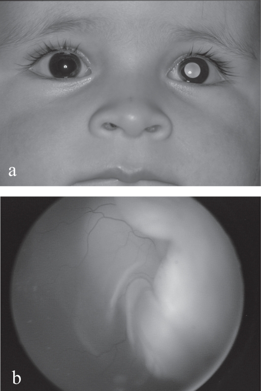
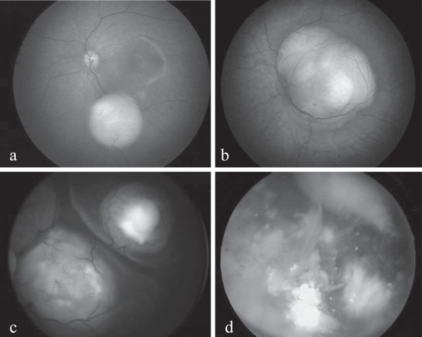
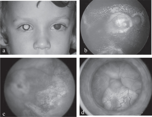
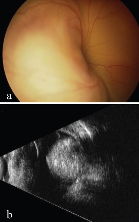
Leukocoria in infants is always a danger signal as retinoblastoma, a malignant retinal tumor, is responsible for half of the cases in this age group. More common signs should also be considered suspicious until proved otherwise, such as strabismus, the second most frequent sign of retinoblastoma. Less frequent manifestations are inflammatory conditions resistant to treatment, hypopyon, orbital cellulitis, hyphema or heterochromia. Other causal pathologies, including persistent hyperplastic primary vitreous (PHPV), Coats' disease, ocular toxocariasis or retinopathy of prematurity, may also manifest the same warning signs and require specialized differential diagnosis. Members of the immediate family circle are most likely to notice the first signs, the general practitioner, pediatrician or general ophthalmologist the first to be consulted. On their attitude will depend the final outcome of this vision and life-threatening disease. Early diagnosis is vital.
Keywords: Coats’ disease; leukocoria; persistent hyperplastic primary vitreous (PHPV); retinoblastoma; strabismus.
Figures
Similar articles
-
Unusual presenting signs of retinoblastoma: a case study.J Ophthalmic Nurs Technol. 1991 May-Jun;10(3):98-102. J Ophthalmic Nurs Technol. 1991. PMID: 2061938
-
Presenting signs of retinoblastoma.J Pediatr. 1998 Mar;132(3 Pt 1):505-8. doi: 10.1016/s0022-3476(98)70028-9. J Pediatr. 1998. PMID: 9544909
-
[Leukokoria in a child: emergency and challenge].Klin Monbl Augenheilkd. 1999 May;214(5):332-5. doi: 10.1055/s-2008-1034807. Klin Monbl Augenheilkd. 1999. PMID: 10420380 Review. French.
-
Leukocoria.2023 Aug 25. In: StatPearls [Internet]. Treasure Island (FL): StatPearls Publishing; 2025 Jan–. 2023 Aug 25. In: StatPearls [Internet]. Treasure Island (FL): StatPearls Publishing; 2025 Jan–. PMID: 32809629 Free Books & Documents.
-
Don't Miss This! Red Flags in the Pediatric Eye Examination: Abnormal Red Reflex.J Binocul Vis Ocul Motil. 2019 Jul-Sep;69(3):106-109. doi: 10.1080/2576117X.2019.1607429. J Binocul Vis Ocul Motil. 2019. PMID: 31329054 Review.
Cited by
-
Hyperoleon masquerading as leukocoria.BMJ Case Rep. 2021 Sep 20;14(9):e246135. doi: 10.1136/bcr-2021-246135. BMJ Case Rep. 2021. PMID: 34544725 Free PMC article. No abstract available.
-
Unilateral Leukocoria in an Infant.Cureus. 2020 Nov 20;12(11):e11596. doi: 10.7759/cureus.11596. Cureus. 2020. PMID: 33364117 Free PMC article.
-
Leukocoria in a 4-year-old boy.Digit J Ophthalmol. 2024 Jun 6;30(2):42-44. doi: 10.5693/djo.02.2024.03.005. eCollection 2024. Digit J Ophthalmol. 2024. PMID: 38962670 Free PMC article.
-
Evaluation of the red reflex: An overview for the pediatrician.World J Methodol. 2021 Sep 20;11(5):263-277. doi: 10.5662/wjm.v11.i5.263. eCollection 2021 Sep 20. World J Methodol. 2021. PMID: 34631483 Free PMC article.
-
Improving Medical Students' Awareness About Retinoblastoma: A Practical Strategy.Clin Ophthalmol. 2022 Jun 7;16:1807-1814. doi: 10.2147/OPTH.S355876. eCollection 2022. Clin Ophthalmol. 2022. PMID: 35698597 Free PMC article.
References
-
- Abramson DH. Second nonocular cancers in retinoblastoma: a unified hypothesis. The Franceschetti Lecture. Ophthalmic Genet. 1999;20:193–204. - PubMed
-
- Abramson DH, Beaverson K, Sangani P, et al. Screening for retinoblastoma: presenting signs as prognosticators of patient and ocular survival. Pediatrics. 2003;112:1248–55. - PubMed
-
- Abramson DH, Frank CM, Susman M, et al. Presenting signs of retinoblastoma. J Pediatr. 1998;132:505–8. - PubMed
-
- Abramson DH, Melson MR, Dunkel IJ, et al. Third (fourth and fifth) nonocular tumors in survivors of retinoblastoma. Ophthalmology. 2001;108:1868–76. - PubMed
LinkOut - more resources
Full Text Sources
