. 2009 Aug 11;4(8):e6528.
doi: 10.1371/journal.pone.0006528.

Merkel cells as putative regulatory cells in skin disorders: an in vitro study

Affiliations

Merkel cells as putative regulatory cells in skin disorders: an in vitro study

Nicholas Boulais et al. PLoS One. .

Abstract

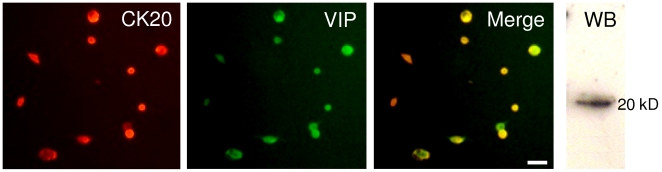
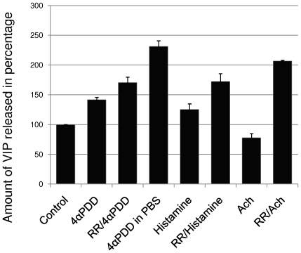
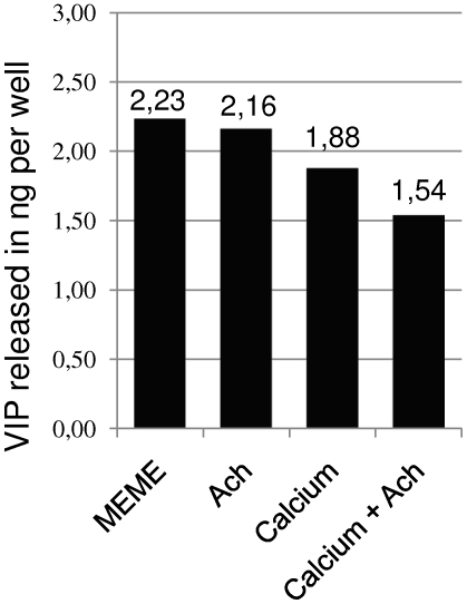
Merkel cells (MCs) are involved in mechanoreception, but several lines of evidence suggest that they may also participate in skin disorders through the release of neuropeptides and hormones. In addition, MC hyperplasias have been reported in inflammatory skin diseases. However, neither proliferation nor reactions to the epidermal environment have been demonstrated. We established a culture model enriched in swine MCs to analyze their proliferative capability and to discover MC survival factors and modulators of MC neuroendocrine properties. In culture, MCs reacted to bFGF by extending outgrowths. Conversely, neurotrophins failed to induce cell spreading, suggesting that they do not act as a growth factor for MCs. For the first time, we provide evidence of proliferation in culture through Ki-67 immunoreactivity. We also found that MCs reacted to histamine or activation of the proton gated/osmoreceptor TRPV4 by releasing vasoactive intestinal peptide (VIP). Since VIP is involved in many pathophysiological processes, its release suggests a putative regulatory role for MCs in skin disorders. Moreover, in contrast to mechanotransduction, neuropeptide exocytosis was Ca(2+)-independent, as inhibition of Ca(2+) channels or culture in the absence of Ca(2+) failed to decrease the amount of VIP released. We conclude that neuropeptide release and neurotransmitter exocytosis may be two distinct pathways that are differentially regulated.


Conflict of interest statement

Competing Interests: The authors have declared that no competing interests exist.

Figures

Figure 1
Figure 1. MC are densely represented in the swine snout.
MCs labeled by CK20 immunostaining (in red) appear abundant in the basal layer of the epidermis of swine snout. Conversely, less than 50 MCs/mm2 were found in human glabrous skin. Nuclei were stained in blue with DAPI. (Scale bar, 50 µm).
Figure 2
Figure 2. Immunofluorescence on selected Merkel cells in culture.
(a) After enrichment, an average of 62% (±3.5) MCs was obtained over 10 experiments as demonstrated by immunostaining using anti-CK20 antibodies and DAPI as a counterstain. In order to define a suitable culture medium for MCs, cells were stimulated by various factors and the morphology of MCs was analyzed by CK20 immunofluorescence. Using DMEM/F12 as a basal medium, neither EGF (20 ng/mL), RA (0.5 µM) or Dexa (1 µM) supported the spreading of MCs. Similarly, neither NGF (100 ng/mL), BDNF (25 ng/mL), NT-3 (25 ng/mL) or the three factors together (b) stimulated the growth of cytoplasmic extensions. (c) Conversely, MCs reacted to bFGF (20 ng/mL), as they extended cytoplasmic outgrowths (arrows), suggesting that this factor acts as a growth factor for MCs. (Scale bar in all pictures, 50 µm).
Figure 3
Figure 3. Electron microscopy analysis confirmed the identity of Merkel cells.
Ultrastructural analyses were carried out on cells from the enriched MC fraction. (a, b) Up to half of the cells presented features characteristic of MCs: a polylobulated nucleus with numerous typical dense-core granules in a clear cytoplasm. (c) The thin membrane distinguishable around the darkest cytoplasmic granules is consistent with neuroendocrine cells. (Scale bar in a, 5 µm; in b, 2 µm; in c, 500 nm).
Figure 4
Figure 4. MCs developed cytoplasmic processes when cultured in the presence of serum.
Strikingly, in these culture conditions, stacking of more than five MCs was observed (a, b) and confirmed by CK20 immunofluorescence (c). Interestingly, this stacking is similar to configurations observed in mechanosensitive areas in the touch pads of rats, as demonstrated by CK20 immunostaining in dissociated epidermal layers (d). The meaning of these alignments to the role of MCs remains to be defined. (Scale bars, 10 µm).
Figure 5
Figure 5. Merkel cells proliferated in culture.
Double immunofluorescence assay using antibodies against CK20 (red) and Ki-67 antigen (green) revealed the ability of MCs to proliferate in vitro. Dividing CK20-positive cells were clearly visible, and all phases of the cell cycle were observed: prophase (a), metaphase (b), anaphase (c, e) and telophase (d, e). (Scale bars, 20 µm).
Figure 6
Figure 6. Merkel cells produced VIP in culture.
In our culture conditions, MCs produced the neuropeptide VIP as demonstrated by double immunofluorescence using antibodies against CK20 (red) and VIP (green). The production of VIP was confirmed by western blot analysis (WB). (Scale bar, 50 µm).
Figure 7
Figure 7. Percentage of VIP released by Merkel cells.
The amount of VIP released in the culture supernatant, assessed by ELISA and expressed as a percentage of the control conditions (DMEM/F12 containing 1 mM Ca2+). Exposure to the TRPV4 agonist 4αPDD (1 µM) or histamine (100 µM) stimulated VIP release from MCs. This release was not inhibited by pre-incubation with the Ca2+ channel inhibitor RR (1 µM), indicating a Ca2+-independent pathway. The marked increase of VIP release obtained following stimulation by 4αPDD in a Ca2+-free buffer confirmed these results. In addition, exposure to the neurotransmitter Ach (10 µM) significantly inhibited the amount of VIP released in the presence of Ca2+. This inhibition was suppressed by pre-incubation with RR (1 µM), suggesting the involvement of Ca2+ channels (n = 4, difference significant at P≤0.025.).
Figure 8
Figure 8. Ca2+ and Ach decreased the release of VIP.
Effect of Ach and Ca2+ on VIP release expressed in ng/well. MCs were cultured in Ca2+-free MEME and then exposed to Ach (100 µM), Ca2+ (1 mM), or both. In the absence of Ca2+, the addition of Ach failed to significantly decrease VIP exocytosis (P>0.1). A lower amount of VIP release was observed in the presence of Ca2+ (1 mM). Finally, the inhibitory effect of Ach on VIP release required extracellular Ca2+ (P≤0.025). These results are consistent with inhibition via Ca2+ channels, probably via muscarinic M2 or M4 Ach receptors.

References

    1. Moll I, Roessler M, Brandner JM, Eispert AC, Houdek P, et al. Human Merkel cells–aspects of cell biology, distribution and functions. Eur J Cell Biol. 2005;84:259–271. - PubMed
    1. Halata Z, Grim M, Bauman KI. Friedrich Sigmund Merkel and his “Merkel cell”, morphology, development, and physiology: review and new results. Anat Rec A Discov Mol Cell Evol Biol. 2003;271:225–239. - PubMed
    1. Boulais N, Misery L. Merkel cells. J Am Acad Dermatol. 2007;57:147–165. - PubMed
    1. Ogawa H. The Merkel cell as a possible mechanoreceptor cell. Prog Neurobiol. 1996;49:317–334. - PubMed
    1. Yamashita Y, Akaike N, Wakamori M, Ikeda I, Ogawa H. Voltage-dependent currents in isolated single Merkel cells of rats. J Physiol. 1992;450:143–162. - PMC - PubMed

Publication types

Substances