. 2008 Jun;2(2):413-24.
doi: 10.2147/opth.s2151.

Age and disease-related structural changes in the retinal pigment epithelium

Affiliations
Free PMC article

Age and disease-related structural changes in the retinal pigment epithelium

Vera L Bonilha. Clin Ophthalmol. 2008 Jun.
Free PMC article

Abstract

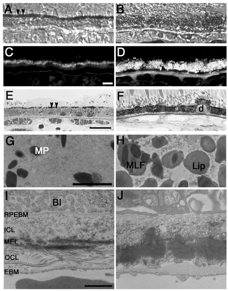
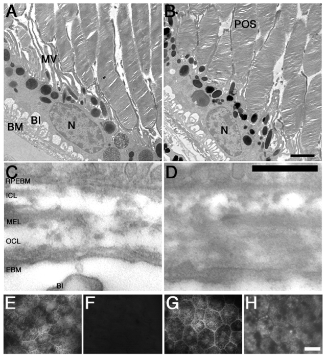
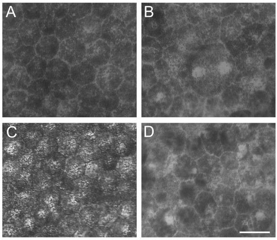
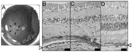
As the retinal pigment epithelium (RPE) ages, a number of structural changes occur, including loss of melanin granules, increase in the density of residual bodies, accumulation of lipofuscin, accumulation of basal deposits on or within Bruch's membrane, formation of drusen (between the basal lamina of the RPE and the inner collagenous layer of Bruch's membrane), thickening of Bruch's membrane, microvilli atrophy and disorganization of the basal infoldings. Although these changes are well known, the basic mechanisms involved in them are frequently poorly understood. These age-related changes progress slowly and vary in severity in different individuals. These changes are also found in age-related macular degeneration (AMD), a late onset disease that severely impacts the RPE, but they are much more pronounced than during normal aging. However, the changes in AMD lead to severe loss of vision. Given the many supporting functions which the RPE serves for the retina, it is important to decipher the age-related changes in this epithelium in order to understand age-related changes in vision.

Keywords: age-related macular degeneration (AMD); aging; ocular disorders; retinal disease; retinal pigment epithelium.


Figures

Figure 1
Figure 1
Age-related changes in human RPE. Observation of the structural differences in RPE from young (23 year-old, A, C, E, G, I) and aged (75 and 88 year-old, B, D, F, H, J) human donors. Aged RPE from human donors displays loss of melanin granules (MP, arrowheads in A, E) and accumulation of the age pigment lipofuscin (Lip) (B, D), as observed by the presence of increased autofluorescent granules when observed on epifluorescence in the green channel (FITC filter: excitation 495 nm/emission 519 nm) in aged RPE (D) when compared to young RPE (C). Additional observation of the aged RPE displayed formation of drusen (D) (between the basal lamina of the RPE and the inner collagenous layer of Bruch’s membrane) (F), thickening of Bruch’s membrane and basal infoldings disorganization (J) when processed and analyzed by electron microscopy. In addition, in the aged RPE cells melanin granules are frequently seen in association with lipofuscin (melanolipofuscin, MLF) granules (H). Young RPE displays melanin pigments on their apical surface (A, E, G) while aged RPE contains mostly lipofuscin granules (B, H). Differential interference contrast microscopy images (A, B). Semi-thin epon sections stained with toluidine blue of young (E) and aged RPE (F) examined in bright field. Abbreviations: BI, basal infoldings; RPEBM, RPE basement membrane; ICL, inner collagenous layer; MEL, middle elastic layer; OCL, outer collagenous layer; EBM, choroidal endothelial cell basement membrane; Bars: (A to D), 10 μm; (E, F), 200 μm; (G, H), 2 μm; (I, J), 1 μm.
Figure 2
Figure 2
Age-related changes in F1 F344BN hybrid rat RPE. Observation of young (3–4 month-old, A) and aged (24–25 month-old, B) F1 F344BN hybrid rats reveals several of the RPE age-related changes previously described. These include: Bruch’s membrane thickening (D), accumulation of residual bodies, and microvilli atrophy (B). In addition, bright-field analysis of aged RPE whole-mounts reveals decrease in RPE density (G) while epifluorescence in the green channel (FITC filter: excitation 495 nm/emission 519 nm) reveals increased lipofuscin accumulation (H) when compared to the young RPE cells (E and F). AD. Transmission electron microscopy. Abbreviations: BI, basal infoldings; MV, microvilli; POS, photorecptor outer segments; RPEBM, RPE basement membrane; ICL, inner collagenous layer; MEL, middle elastic layer; OCL, outer collagenous layer; EBM, choroidal endothelial cell basement membrane; Bars: (A and B), 1 μm; (C and D), 2 μm; and (C to F), 200 μm.
Figure 3
Figure 3
Age-related changes in RPE density. Bright-field micrographs of RPE whole- mounts from both young (A and B) and aged (C and D) donor eyes. Observations were carried out both in the fovea (A and C) and periphery (B and D) of the eyes. Foveal RPE cells are smaller and more homogeneous than the peripheral RPE cells independent of the age of the donor. Bas, 200 μm.
Figure 4
Figure 4
RPE cell loss in geographic atrophy. Gross photomicrography of a postmortem eye from an AMD donor with geographic atrophy (GA) (A). Arrows indicate the edges of GA; inset indicates the region cut and processed for transmission electron microscopy. Semi-thin epon sections stained with toluidine blue of this region demonstrates extensive RPE, photoreceptors and choroids atrophy in the GA region (B) while the edge of GA displays more RPE cells and the presence of some photoreceptor outer segments (C). In the region outside of the GA the RPE layer is continuous and the photoreceptors’ inner and outer segments can be observed (D). Arrowheads point to Bruch’s membrane. Debris accumulation is observed underneath the RPE cells in all regions observed. Bars 200 μm.

References

    1. Anderson DH, Mullins RF, Hageman GS, et al. A role for local inflammation in the formation of drusen in the aging eye. Am J Ophthalmol. 2002;134:411–30. - PubMed
    1. Arnold JJ, Quaranta M, Soubrane G, et al. Indocyanine green angiography of drusen. Am J Ophthalmol. 1997;124:344–56. - PubMed
    1. Bando H, Shadrach KG, Rayborn ME, et al. Clathrin and adaptin accumulation in drusen, Bruch’s membrane and choroid in AMD and non-AMD donor eyes. Exp Eye Res. 2007;84:135–42. - PubMed
    1. Baynes JW. The role of AGEs in aging: causation or correlation. Exp Gerontol. 2001;36:1527–37. - PubMed
    1. Beatty S, Koh H-H, Phil M, et al. The role of oxidative stress in the pathogenesis of age-related macular degeneration. Surv Ophthalmol. 2000;45:115–34. - PubMed