

Optical coherence tomography findings in paraneoplastic pseudovitelliform lesions in melanoma-associated retinopathy
- PMID: 19668738
- PMCID: PMC2693986
- DOI: 10.2147/opth.s2282
Optical coherence tomography findings in paraneoplastic pseudovitelliform lesions in melanoma-associated retinopathy
Abstract
Purpose: To report an unusual case of paraneoplastic pseudovitelliform lesions associated with melanoma-associated retinopathy (MAR).
Design: Observational case report.
Methods: Retrospective review of the ophthalmic examination, fundus photography, fluorescein angiography, electroretinogram (ERG), and optical coherence tomography (OCT) of a patient with MAR.
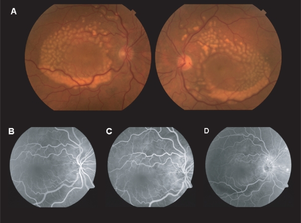
Results: A 65-year-old Caucasian man with a two-year history of metastatic melanoma was referred for evaluation of a six-month history of nyctalopia. Funduscopic examination in both eyes revealed multiple, creamy, yellow, pseudovitelliform lesions in the posterior pole, varying in size from 100-500 mum, at the level of the outer retinal/retinal pigment epithelium (RPE) junction, coalescing along the inferior portion, with overlying macular neurosensory detachments. OCT showed bilateral macular neurosensory detachments with multiple small areas of high reflectivity at the level of the outer retinal/RPE junction. ERG demonstrated a selective loss of the b-wave and a normal a-wave under dark adapted, scotopic conditions.
Conclusion: Clinicians should be aware of this atypical presentation of MAR that may include pseudovitelliform retinal findings.
Keywords: cancer-associated retinopathy; melanoma-associated retinopathy; optical coherence tomography; paraneoplastic pseudovitelliform retinopathy; paraneoplastic syndrome.
Figures

References
-
- Borkowski LM, Grover S, Fishman GA, et al. Retinal findings in melanoma-associated retinopathy. Am J Ophthalmol. 2001;132:273–5. - PubMed
-
- Palmowski AM, Haus AH, Pfohler C, et al. Bilateral multifocal chorioretinopathy in a woman with cutaneous malignant melanoma. Arch Ophthalmol. 2002;120:1756–61. - PubMed
-
- Potter MJ, Thirkill CE, Dam OM, et al. Clinical and immunocytochemical findings in a case of melanoma-associated retinopathy. Ophthalmology. 1999;106:2121–5. - PubMed
-
- Sotodeh M, Paridaens D, Keunen J, et al. Paraneoplastic vitelliform retinopathy associated with cutaneous or uveal melanoma and metastases. Klin Monatsbl Augenheilkd. 2005;222:910–14. - PubMed
-
- Zacks DN, Pinnolis MK, Berson EL, et al. Melanoma-associated retinopathy and recurrent exudative retinal detachments in a patient with choroidal melanoma. Am J Ophthalmol. 2001;132:578–81. - PubMed
Publication types
LinkOut - more resources
Full Text Sources
