

Sudden unilateral visual loss after autologous fat injection into the nasolabial fold
- PMID: 19668775
- PMCID: PMC2694002
Sudden unilateral visual loss after autologous fat injection into the nasolabial fold
Abstract
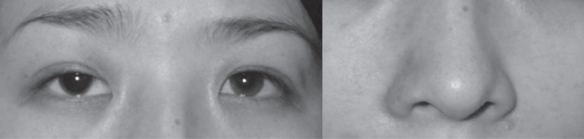
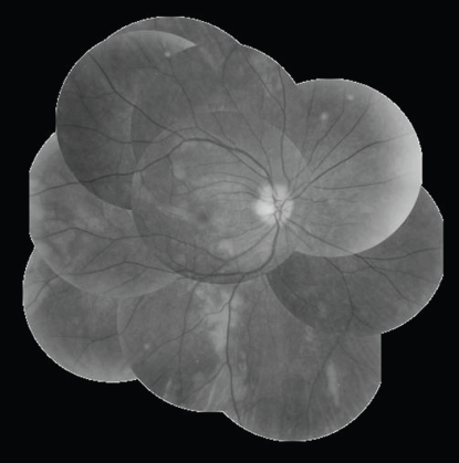
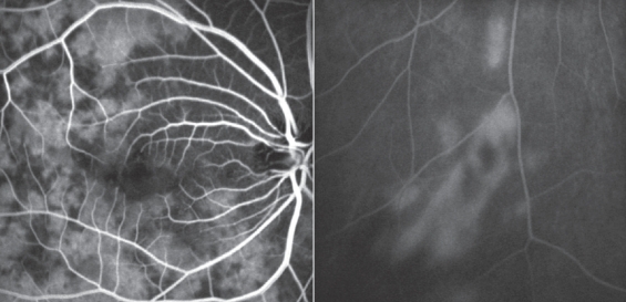
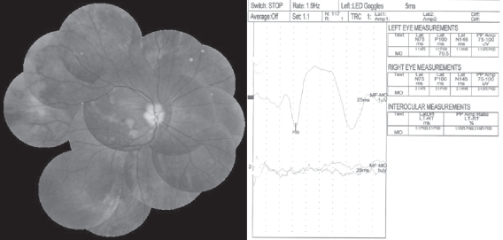
A 27-year-old female presented with sudden visual loss of her right eye after receiving an autologous fat injection into the right nasolabial fold. Fundus examination of the right eye showed multiple whitish patchy lesions with macular edema. Fluorescein angiogram showed deterioration of choroidal circulation with patchy choroidal filling and arm-to-retina circulation time and retinal arteriovenous passage time were delayed to 30 seconds and 20 seconds, respectively. There was no response in flash visual evoked potential (VEP). High dose steroid therapy (methylprednisolone 1 g/day/i.v.) was done and about 2 weeks later, the disc edema subsided and retinal arteriovenous passage time of fluorescein angiogram was normalized but there was no improvement in visual acuity. Absence of a cherry red spot, deterioration of choroidal circulation with patchy choroidal fillings seen in fluorescein angiogram, and no response in flash VEP suggests multiple choroidal infarction due to perfusion defect of the short posterior ciliary artery. The autologous fat injected is thought to have entered the dorsal nasal artery and the retrograde migration of the emboli to the ophthalmic artery might have caused the multiple occlusions of the short posterior ciliary artery.
Keywords: autologous fat injection; ciliary artery occlusion; ischemic optic neuropathy.
Figures
References
-
- Caplan LR. Brain embolism, revisited. Neurology. 1993;43:1281–7. - PubMed
-
- Cheney ML, Blair PA. Blindness as a complication of rhinoplasty. Arch Otolaryngol Head Neck Surg. 1987;113:768–9. - PubMed
-
- Danesh-Meyer HV, Savino PJ, Sergott RC. Case reports and small case series: ocular and cerebral ischemia following facial injection of autologous fat. Arch Ophthalmol. 2001;119:777–8. - PubMed
-
- Dreizen NG, Framm L. Sudden unilateral visual loss after autologous fat injection into the glabellar area. Am J Ophthalmol. 1989;107:85–7. - PubMed
-
- Egido JA, Arroyo R, Marcos A, et al. Middle cerebral artery embolism and unilateral visual loss after autologous fat injection into the glabellar area. Stroke. 1993;24:615–16. - PubMed
Publication types
LinkOut - more resources
Full Text Sources
