

Extranodal marginal zone lymphoma of the dura mater with IgH/MALT1 translocation and review of literature
- PMID: 19669212
- PMCID: PMC2713483
- DOI: 10.1007/s12308-008-0005-9
Extranodal marginal zone lymphoma of the dura mater with IgH/MALT1 translocation and review of literature
Abstract
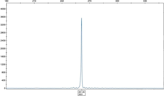
Primary central nervous system lymphoma (PCNSL) is an extranodal non-Hodgkin lymphoma involving brain, intraocular structures and spinal cord, without evidence of systemic disease. The majority of PCNSLs are diffuse large B-cell type. We encountered a rare case of primary dural marginal zone lymphoma of mucosa-associated lymphoid tissue (MALT) with extension into the brain in a 59-year-old man. A magnetic resonance imaging scan showed a 22-mm tumor located in the left posterior temporal lobe extending from the dura. Histopathology revealed a lymphoplasmacytic infiltration of the dura and the brain parenchyma in a perivascular pattern. Immunohistochemical and in situ hybridization studies showed a B-cell phenotype with kappa light chain restriction. Fluorescent in situ hybridization study showed a t(14;18)(q32;q21) with immunoglobulin heavy-chain/MALT1 fusion. The molecular study for immunoglobulin heavy-chain gene rearrangement by polymerase chain reaction showed a clonal gene rearrangement.
Figures

Similar articles
-
Primary cutaneous marginal zone B-cell lymphoma may exhibit both the t(14;18)(q32;q21) IGH/BCL2 and the t(14;18)(q32;q21) IGH/MALT1 translocation: an indicator for clonal transformation towards higher-grade B-cell lymphoma?Am J Dermatopathol. 2007 Jun;29(3):231-6. doi: 10.1097/DAD.0b013e31804795a6. Am J Dermatopathol. 2007. PMID: 17519619
-
The t(14;18)(q32;q21)/IGH-MALT1 translocation in gastrointestinal extranodal marginal zone lymphoma of mucosa-associated lymphoid tissue (MALT lymphoma).Histopathology. 2014 May;64(6):791-8. doi: 10.1111/his.12327. Epub 2014 Jan 10. Histopathology. 2014. PMID: 24236896
-
T(14;18)(q32;q21) involving IGH and MALT1 is a frequent chromosomal aberration in MALT lymphoma.Blood. 2003 Mar 15;101(6):2335-9. doi: 10.1182/blood-2002-09-2963. Epub 2002 Oct 24. Blood. 2003. PMID: 12406890
-
MALT lymphoma involving the kidney: a report of 10 cases and review of the literature.Am J Clin Pathol. 2007 Sep;128(3):464-73. doi: 10.1309/0T2UKUKV91W3QR6W. Am J Clin Pathol. 2007. PMID: 17709321 Review.
-
Anti-apoptotic action of API2-MALT1 fusion protein involved in t(11;18)(q21;q21) MALT lymphoma.Apoptosis. 2005 Jan;10(1):25-34. doi: 10.1007/s10495-005-6059-6. Apoptosis. 2005. PMID: 15711920 Review.
Cited by
-
Dural MALT lymphoma with disseminated disease.Hematol Rep. 2010 Jan 26;2(1):e10. doi: 10.4081/hr.2010.e10. Epub 2010 Dec 3. Hematol Rep. 2010. PMID: 22184513 Free PMC article.
-
A Rare Case of Composite Dural Extranodal Marginal Zone Lymphoma and Chronic Lymphocytic Leukemia/Small Lymphocytic Lymphoma.Front Neurol. 2018 Apr 24;9:267. doi: 10.3389/fneur.2018.00267. eCollection 2018. Front Neurol. 2018. PMID: 29740389 Free PMC article. Review.
-
Recent Advances in the Genetic of MALT Lymphomas.Cancers (Basel). 2021 Dec 30;14(1):176. doi: 10.3390/cancers14010176. Cancers (Basel). 2021. PMID: 35008340 Free PMC article. Review.
-
A 44-year old male with right-sided facial numbness. Dura-associated extranodal marginal zone B cell lymphoma (MALT lymphoma).Brain Pathol. 2015 Jan;25(1):113-4. doi: 10.1111/bpa.12234. Brain Pathol. 2015. PMID: 25521183 Free PMC article. No abstract available.
-
Extranodal Marginal Zone B-Cell Lymphoma of Mucosa-Associated Tissue Type Involving the Dura.Cancer Res Treat. 2016 Apr;48(2):859-63. doi: 10.4143/crt.2014.334. Epub 2015 Jul 17. Cancer Res Treat. 2016. PMID: 26194368 Free PMC article.
References
-
- Primary brain tumors in the United States 1995–1999: statistical report. CBTRUS: Chicago, IL; 2002–2003.
-
- Kleihues P, Cavenee WK. World Health Organization classification of tumors: pathology and genetics: tumors of the nervous system. Lyon: IARC; 2000.
LinkOut - more resources
Full Text Sources
Research Materials
