. 2008 Apr;34(1-2):197-212.
doi: 10.1007/s10867-008-9114-z. Epub 2008 Oct 4.

Regulation of the F1F0-ATP synthase rotary nanomotor in its monomeric-bacterial and dimeric-mitochondrial forms

Affiliations

Regulation of the F1F0-ATP synthase rotary nanomotor in its monomeric-bacterial and dimeric-mitochondrial forms

José J García-Trejo et al. J Biol Phys. 2008 Apr.

Abstract

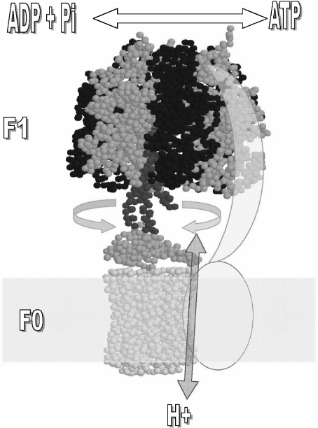
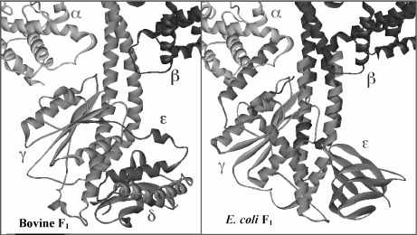
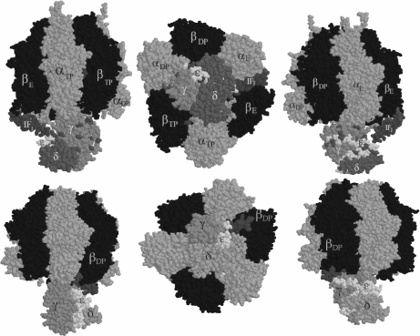
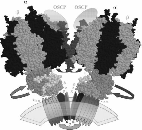
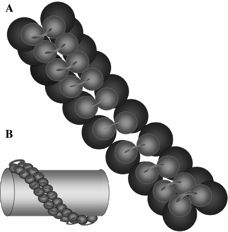
The F(1)F(0)-adenosine triphosphate (ATP) synthase rotational motor synthesizes most of the ATP required for living from adenosine diphosphate, Pi, and a proton electrochemical gradient across energy-transducing membranes of bacteria, chloroplasts, and mitochondria. However, as a reversible nanomotor, it also hydrolyzes ATP during de-energized conditions in all energy-transducing systems. Thus, different subunits and mechanisms have emerged in nature to control the intrinsic rotation of the enzyme to favor the ATP synthase activity over its opposite and commonly wasteful ATPase turnover. Recent advances in the structural analysis of the bacterial and mitochondrial ATP synthases are summarized to review the distribution and mechanism of the subunits that are part of the central rotor and regulate its gyration. In eubacteria, the epsilon subunit works as a ratchet to favor the rotation of the central stalk in the ATP synthase direction by extending and contracting two alpha-helixes of its C-terminal side and also by binding ATP with low affinity in thermophilic bacteria. On the other hand, in bovine heart mitochondria, the so-called inhibitor protein (IF(1)) interferes with the intrinsic rotational mechanism of the central gamma subunit and with the opening and closing of the catalytic beta-subunits to inhibit its ATPase activity. Besides its inhibitory role, the IF(1) protein also promotes the dimerization of the bovine and rat mitochondrial enzymes, albeit it is not essential for dimerization of the yeast F(1)F(0) mitochondrial complex. High-resolution electron microscopy of the dimeric enzyme in its bovine and yeast forms shows a conical shape that is compatible with the role of the ATP synthase dimer in the formation of tubular the cristae membrane of mitochondria after further oligomerization. Dimerization of the mitochondrial ATP synthase diminishes the rotational drag of the central rotor that would decrease the coupling efficiency between rotation of the central stalk and ATP synthesis taking place at the F(1) portion. In addition, F(1)F(0) dimerization and its further oligomerization also increase the stability of the enzyme to natural or experimentally induced destabilizing conditions.


Figures

Fig. 1
Fig. 1
Rotor–stator subunit distribution in the mitochondrial F1F0-ATP synthase. Only the core subunits present in bacteria and mitochondria are shown for simplicity. Rotating subunits are shown in red (γ), orange (ε), and yellow (ring of c subunits) whereas static subunits are in dark blue (β), blue (α), and cyan (subunits a and b). The arrows indicate the reversible rotation of γ–ε–c subunits relative to α and β catalytic subunits of F1 that takes place during ATP synthesis (“clockwise” or right direction) and hydrolysis (“counterclockwise” or left direction). Bidirectional proton flow at the c-ring–sub a interface occurs associated with the gyration of the rotor as indicated by the red arrow. The second-stalk structure is simplified as two cyan subunits (a and b) that work as stator to anchor the catalytic α3β3 to the membranous a subunit. Image is generated in RasMol 2.6 from the mitochondrial F1F0 structure of S. cerevisiae (PDB code 1Q01) and edited as shown
Fig. 2
Fig. 2
Comparison of the bovine (left) and E. coli (right) F1F0-ATP synthases at the central stalk domain. Crystallographic structures of MF1 and EF1 central stalks are shown in the same orientation. Homologous subunits are drawn in the same color, γ (blue), ε subunits (green). For clarity, only one α subunit (red) and one β subunit (yellow) are shown. The structure shown on the right is a composite of the E. coli γ–ε structure [18] and the bovine MF1 structure [19], constructed by aligning segments of γ present in both structures. Segments of MF1 γ subunit are shown in darker blue, and those of E. coli γ subunit are shown in lighter blue. This figure was modified from an original courtesy of Dr. Andrew J.W. Rodgers
Fig. 3
Fig. 3
Model and crystal structures of the F1–IF1 complex from bovine heart mitochondria. Top three panels, our model: we positioned the IF1 N-terminal domain at an entrance-binding site (αE–βE interface) at about 12-Å cross-linking distance from γ and ε subunits as we found [44]. From the side (left and right) and “bottom” (center) views, it was clearly shown and proposed for the first time that IF1 is close enough to the rotor of the enzyme to block gyration of the central stalk as part of its inhibitory mechanism [44]. Bottom panels, the crystal structure from the F1–IF1 crystal with a nondimerizing fragment of IF1 [21]: the same IF1 N-terminal side was resolved and observed actually bound to the γ subunit at an αDP–βDP interface [21, 22]. The top structure depicts the entrance site of IF1, whereas the bottom structure shows the final inhibited structure where IF1 is locked into the same αE–βE interface that became αDP–βDP after two counterclockwise 120° gyration steps (shift from top to bottom panels). IF1 therefore inhibits rotation of the central stalk and the opening and closing conformational changes of a single catalytic interface
Fig. 4
Fig. 4
Model of the dimeric-mitochondrial ATP synthase: possible localization of the IF1 protein and its movements to allow rotation of the central stalk during ATP synthesis. The model depicts the overall shape of the dimeric ATP synthase molecule that we observed for the bovine mitochondrial enzyme [54]. The dimeric interface involves F0 subunits (e and g) and two protein bridges, one at the F0–F0 side of unknown composition (question mark) and another at the F1–F1 interface where the second stalks (not shown for clarity) and the IF1 protein (red) are likely to be located. The C-terminal side of the IF1 molecule is assumed to cross the dimer interface and to stabilize the dimer by interacting with subunits OSCP [65] and possibly subunits of the second stalk. The N-terminal inhibitory domain that in the absence of the proton gradient blocks rotation of the central stalk by entering at an α–β–γ interface (Fig. 2) is removed from this position and exposed into the media after establishment of a transmembrane proton gradient, thus allowing rotation of the central stalk during ATP synthesis. The F1 structures were constructed from the bovine F1-DCCD coordinates available (PDF code 1E79)
Fig. 5
Fig. 5
Possible arrangement of the ATP synthase helical polymer that wraps and gives shape to the mitochondrial tubular cristae. a Inner view of the polymer from the interior of the cristae (membrane is transparent); the green spheres represent the F0 channels connected by the protein bridge we observed [54]; the blue spheres are the F1 heads. b The ATP synthase polymer is depicted from the outer surface of a single cristae (yellow). The F1 particles are in blue and connected with a protein bridge that could be composed by the IF1 and second-stalk subunits. The original model is from Allen et al. [75]

Similar articles

Cited by

References

    1. {'text': '', 'ref_index': 1, 'ids': [{'type': 'PubMed', 'value': '13734097', 'is_inner': True, 'url': 'https://pubmed.ncbi.nlm.nih.gov/13734097/'}]}
    2. Penefsky, H.S., Pullman, M.E., Datta, A., Racker, E.: Partial resolution of the enzymes catalyzing oxidative phosphorylation. II. Participation of a soluble adenosine triphosphatase in oxidative phosphorylation. J. Biol. Chem. 235, 3330–3336 (1960) - PubMed
    1. {'text': '', 'ref_index': 1, 'ids': [{'type': 'DOI', 'value': '10.1007/BF00831537', 'is_inner': False, 'url': 'https://doi.org/10.1007/bf00831537'}, {'type': 'PubMed', 'value': '7721724', 'is_inner': True, 'url': 'https://pubmed.ncbi.nlm.nih.gov/7721724/'}]}
    2. Souid, A.K., Penefsky, H.S.: Mechanism of ATP synthesis by mitochondrial ATP synthase from beef heart. J. Bioenerg. Biomembranes 26, 627–630 (1994). doi:10.1007/BF00831537 - PubMed
    1. {'text': '', 'ref_index': 1, 'ids': [{'type': 'DOI', 'value': '10.1111/j.1432-1033.1997.00622.x', 'is_inner': False, 'url': 'https://doi.org/10.1111/j.1432-1033.1997.00622.x'}, {'type': 'PubMed', 'value': '9370375', 'is_inner': True, 'url': 'https://pubmed.ncbi.nlm.nih.gov/9370375/'}]}
    2. García, J.J., Gómez-Puyou, A., Maldonado, E., Tuena de Gómez-Puyou, M.: Acceleration of unisite catalysis of mitochondrial F1-adenosinetriphosphatase by ATP, ADP and pyrophosphate—hydrolysis and release of the previously bound [gamma-32P]ATP. Eur. J. Biochem. 249(2), 622–229 (1997). doi:10.1111/j.1432-1033.1997.00622.x - PubMed
    1. {'text': '', 'ref_index': 1, 'ids': [{'type': 'DOI', 'value': '10.1074/jbc.273.26.15940', 'is_inner': False, 'url': 'https://doi.org/10.1074/jbc.273.26.15940'}, {'type': 'PubMed', 'value': '9632641', 'is_inner': True, 'url': 'https://pubmed.ncbi.nlm.nih.gov/9632641/'}]}
    2. García, J.J., Capaldi, R.A.: Unisite catalysis without rotation of the gamma-epsilon domain in Escherichia coli F1-ATPase. J. Biol. Chem. 273(26), 15940–15945 (1998). doi:10.1074/jbc.273.26.15940 - PubMed
    1. None
    2. García, J.J.: The F0F1-ATP synthase: binding energy, coupling and rotational catalysis. In: Pandalai, S.G. (ed.) Recent Research Developments in Bioenergetics, p. 41. Transworld Research Network, Trivandrum (2000)

LinkOut - more resources