. 2009 Nov;118(5):605-16.
doi: 10.1007/s00401-009-0581-5. Epub 2009 Aug 9.

Abundant FUS-immunoreactive pathology in neuronal intermediate filament inclusion disease

Affiliations

Abundant FUS-immunoreactive pathology in neuronal intermediate filament inclusion disease

Manuela Neumann et al. Acta Neuropathol. 2009 Nov.

Abstract

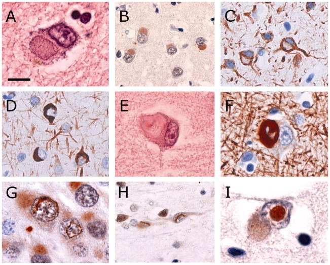
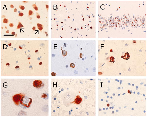
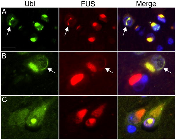
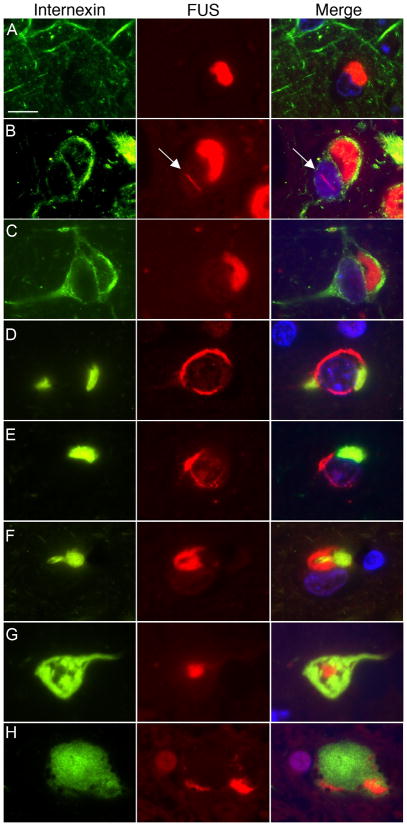
Neuronal intermediate filament inclusion disease (NIFID) is an uncommon neurodegenerative condition that typically presents as early-onset, sporadic frontotemporal dementia (FTD), associated with a pyramidal and/or extrapyramidal movement disorder. The neuropathology is characterized by frontotemporal lobar degeneration with neuronal inclusions that are immunoreactive for all class IV intermediate filaments (IF), light, medium and heavy neurofilament subunits and alpha-internexin. However, not all the inclusions in NIFID are IF-positive and the primary molecular defect remains uncertain. Mutations in the gene encoding the fused in sarcoma (FUS) protein have recently been identified as a cause of familial amyotrophic lateral sclerosis (ALS). Because of the recognized clinical, genetic and pathological overlap between FTD and ALS, we investigated the possible role of FUS in NIFID. We found abnormal intracellular accumulation of FUS to be a consistent feature of our NIFID cases (n = 5). More neuronal inclusions were labeled using FUS immunohistochemistry than for IF. Several types of inclusions were consistently FUS-positive but IF-negative, including neuronal intranuclear inclusions and glial cytoplasmic inclusions. Double-label immunofluorescence confirmed that many cells had only FUS-positive inclusions and that all cells with IF-positive inclusions also contained pathological FUS. No mutation in the FUS gene was identified in a single case with DNA available. These findings suggest that FUS may play an important role in the pathogenesis of NIFID.


Figures

Fig. 1
Fig. 1
Types of neuronal inclusions found in NIFID. Pick body-like inclusions are well-defined, round or oval, slightly eosinophilic or basophilic (a) and show consistent, but often weak, immunoreactivity for ubiquitin (b). They usually do not immunostain for class IV intermediate filaments (IFs), however, many neurons that contain a Pick body-like inclusion show strong, diffuse immunoreactivity for IF in the surrounding cytoplasm (c). Other morphological types of neuronal cytoplasmic inclusions include thin crescents, annular rings and tangle-like inclusions (d). Hyaline conglomerate inclusions appear as irregular, multilobulated masses with a glassy, filamentous appearance and often have a dense, brightly eosinophilic core (e). The filamentous component is strongly immunoreactive for IFs, however the core is often unstained (f). Vermiform neuronal intranuclear inclusions are most frequent in dentate granule cells (g) but also found in pyramidal neurons of the neocortex, hippocampus and some subcortical regions (h). Round, eosinophilic, ubiquitin-immunoreactive neuronal intranuclear inclusions are only present in some cases (i). Neurons with either type of intranuclear inclusion often also contain a Pick body-like cytoplasmic inclusion (g, i). Hematoxylin and eosin (a, e), ubiquitin (b, gi), α-internexin (c, d) and phosphorylated neurofilament (f) immunohistochemistry. Scale bar = 8 μm (a, e, g, i); 25 μm (b); 20 μm (c, d, h); 15 μm (f).
Fig. 2
Fig. 2
FUS immunohistochemistry in NIFID. The normal physiological staining pattern of FUS is demonstrated with higher concentrations of primary antibody and consists of strong immunoreactivity of neuronal nuclei, weaker but consistent staining of neuronal cytoplasm and more variable reactivity of glial nuclei (a). Neurons with FUS-immunoreactive (FUS-ir) inclusions (arrows) still retain some normal physiological FUS staining (a). Abundant FUS-ir pathology is present in all affected brain regions, including the neocortex (b) and hippocampus (c). FUS-ir Pick body-like inclusions (ad), crescentic, annular and tangle-like neuronal cytoplasmic inclusions (e) are numerous. FUS immunohistochemistry occasionally labels structures recognizable as hyaline conglomerate inclusions (f, inferior olive). Many neurons with a FUS-ir vermiform intranuclear inclusion also contain a Pick body-like cytoplasmic inclusion (g). Aggregates of coarse cytoplasmic granules are common in many subcortical regions, including lower motor neurons of the spinal cord (h). Glial cytoplasmic inclusions are common in the cerebral white matter (i). FUS immunohistochemistry with primary antibody from Sigma-Aldrich (HPA008784 vs. epitope aa 86–213) (ai) or Bethyl Laboratories (A300–302A vs. epitope aa 1–50) (c, insert). Scale bar = 25 μm (a, e); 60 μm (b, c); 30 μm (d); 20 μm (f, h, i); 8 μm (g).
Fig. 3
Fig. 3
Double-label immunofluorescence for ubiquitin (green) and FUS (red) in NIFID. Merged images show cell nuclei stained with DAPI (blue). Ubiquitin-positive neuronal cytoplasmic inclusions (NCIs) and vermiform neuronal intranuclear inclusions (NIIs, arrows) always show strong labeling for FUS (a, b). Occasionally, FUS-positive NCIs do not label for ubiquitin (red NCI in merged image, b). A round NII showing colocalization of ubiquitin and FUS. Scale bar = 15 μm (a, b); 10 μm (c).
Fig. 4
Fig. 4
Double-label immunofluorescence for α-internexin (green) and FUS (red) in NIFID. Merged images show cell nuclei stained with DAPI (blue). Many neuronal cytoplasmic inclusions (NCIs) only label for FUS (ac). A subset of neurons with NCI that are only FUS-positive show strong diffuse cytoplasmic staining for α-internexin (b, c). Vermiform neuronal intranuclear inclusions (arrow) only label for FUS (b). Neurons with compact α-internexin-positive inclusions always show additional FUS pathology (dh). However, note that each marker labels separate components of the inclusions, with only marginal overlap. Hyaline conglomerate inclusions (g, h) are composed mainly of α-internexin but always have at least a small FUS-immunoreactive component, either as a central dot (g), or at the periphery of inclusion (h). Scale bar = 10 μm.

Comment in

References

    1. Al Chalabi A, Miller CC. Neurofilaments and neurological disease. Bioessays. 2003;25:346–355. - PubMed
    1. Aman P, Panagopoulos I, Lassen C, et al. Expression patterns of the human sarcoma-associated genes FUS and EWS and the genomic structure of FUS. Genomics. 1996;37:1–8. - PubMed
    1. Andersson MK, Stahlberg A, Arvidsson Y, et al. The multifunctional FUS, EWS, and TAF15 proto-oncoproteins show cell type-specific expression patterns and involvement in cell spreading and stress response. BMC Cell Biol. 2008;9:37. - PMC - PubMed
    1. Baechtold H, Kuroda M, Sok J, Ron D, Lopez BS, Akhmedov AT. Human 75-kDa DNA-pairing protein is identical to the pro-oncoprotein TLD/FUS and is able to promote D-loop formation. J Biol Chem. 1999;274:34337–34342. - PubMed
    1. Bertrand P, Akhmedov AT, Delacote F, Durrbach A, Lopez BS. Human POMp75 is identified as the pro-oncoprotein TLS/FUS: both POMp75 and POMp100 DNA homologous pairing activities are associated with cell proliferation. Oncogene. 1999;18:4515–4521. - PubMed

Publication types

MeSH terms