Review
. 2009 Oct;282(4):329-50.
doi: 10.1007/s00438-009-0473-3. Epub 2009 Aug 11.

Large Arf1 guanine nucleotide exchange factors: evolution, domain structure, and roles in membrane trafficking and human disease

Affiliations
Review

Large Arf1 guanine nucleotide exchange factors: evolution, domain structure, and roles in membrane trafficking and human disease

Quynh Trang Bui et al. Mol Genet Genomics. 2009 Oct.

Abstract

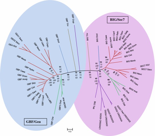
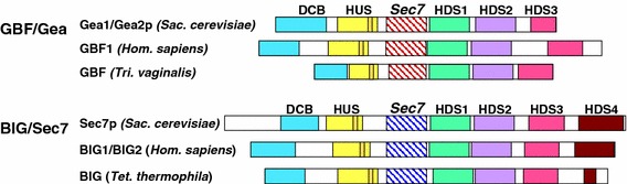
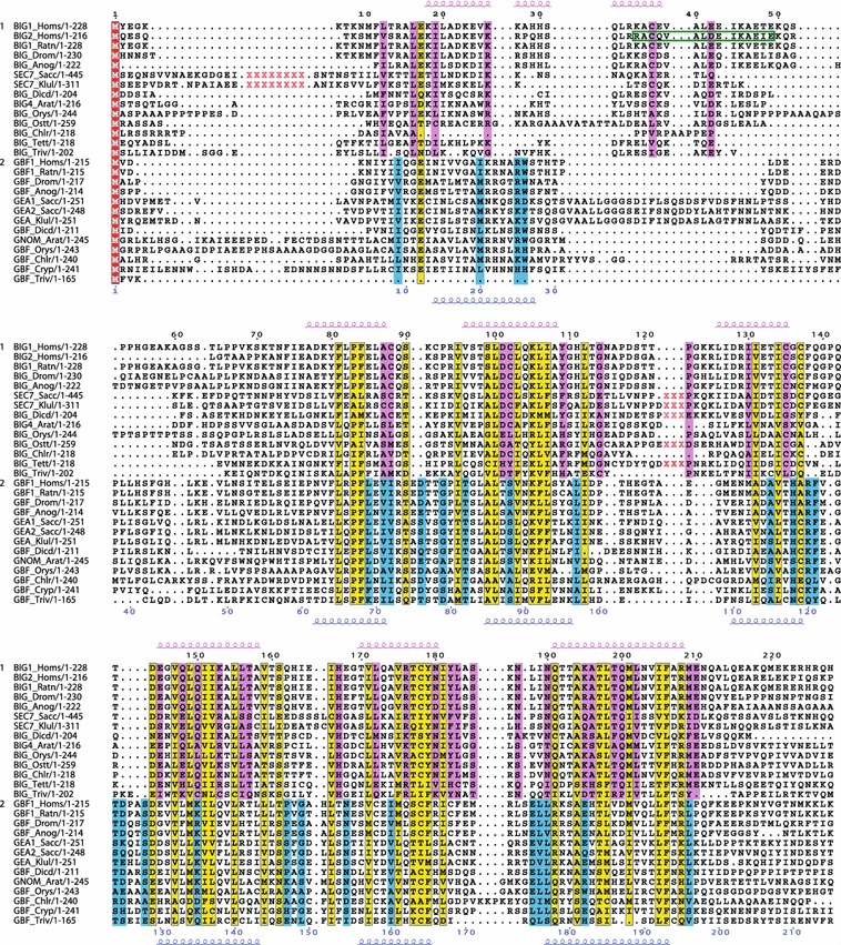
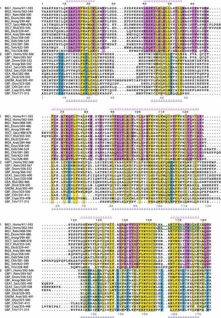
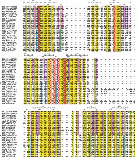
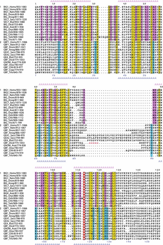
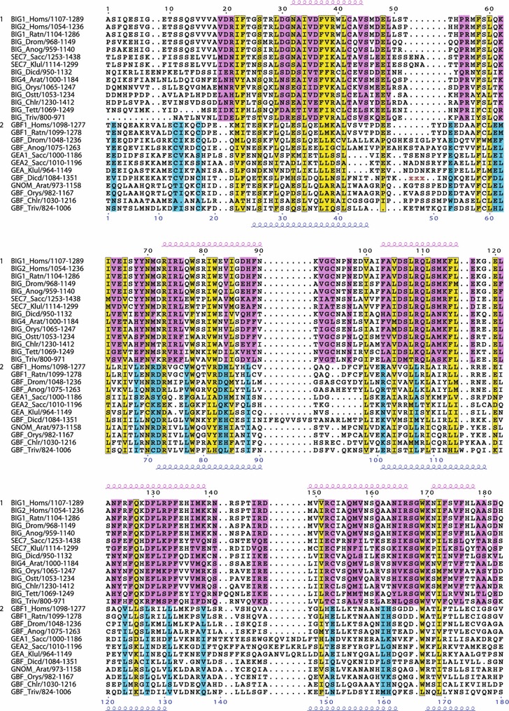
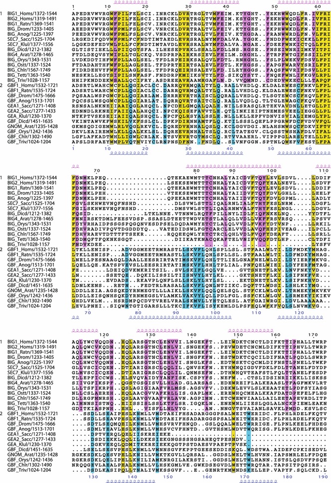
The Sec7 domain ADP-ribosylation factor (Arf) guanine nucleotide exchange factors (GEFs) are found in all eukaryotes, and are involved in membrane remodeling processes throughout the cell. This review is focused on members of the GBF/Gea and BIG/Sec7 subfamilies of Arf GEFs, all of which use the class I Arf proteins (Arf1-3) as substrates, and play a fundamental role in trafficking in the endoplasmic reticulum (ER)-Golgi and endosomal membrane systems. Members of the GBF/Gea and BIG/Sec7 subfamilies are large proteins on the order of 200 kDa, and they possess multiple homology domains. Phylogenetic analyses indicate that both of these subfamilies of Arf GEFs have members in at least five out of the six eukaryotic supergroups, and hence were likely present very early in eukaryotic evolution. The homology domains of the large Arf1 GEFs play important functional roles, and are involved in interactions with numerous protein partners. The large Arf1 GEFs have been implicated in several human diseases. They are crucial host factors for the replication of several viral pathogens, including poliovirus, coxsackievirus, mouse hepatitis coronavirus, and hepatitis C virus. Mutations in the BIG2 Arf1 GEF have been linked to autosomal recessive periventricular heterotopia, a disorder of neuronal migration that leads to severe malformation of the cerebral cortex. Understanding the roles of the Arf1 GEFs in membrane dynamics is crucial to a full understanding of trafficking in the secretory and endosomal pathways, which in turn will provide essential insights into human diseases that arise from misregulation of these pathways.
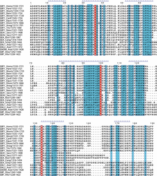

Figures

Fig. 1
Fig. 1
Unrooted phylogenetic tree of the BIG/Sec7 and GBF/Gea Arf GEFs using the full-length sequence of each protein. Three separate analyses were performed using the programs MrBayes (Bayesian inference), PAUP (maximum parsimony), and PhyML (maximum likelihood). Bootstrap percentages are shown for each, with results using MrBayes in bold, PAUP in italics, and a PhyML in normal text. The sequences used for the analysis are shown in Tables 1 and 2. Branches of the tree are color coded to indicate the eukaryotic supergroup that each sequence belongs to: Opisthokonta (red), Archaeplastidia (green), Amoebozoa (blue), Chromalveolata (violet), and Excavata (beige)
Fig. 2
Fig. 2
Domain structure of the BIG/Sec7 and GBF/Gea Arf GEFs. Positions of the homology domains are shown for representative sequences of the Opisthokonta supergroup (Saccharomyces cerevisiae, Homo sapiens), and of the Excavata and Chromalveolata supergroups (Trichomonas vaginalis, Tetrahymena thermophila); positions of domains and sizes of proteins are drawn to scale. The only homology domain unique to one subfamily is the BIG/Sec7 HDS4 sequence; only a portion of the region shown in Fig. 10 is conserved in the Chromalveolata members that we analyzed (amino acid residues 1,600–1,665 of the Tetrahymena thermophila BIG protein, corresponding to amino acids 1,675–1,740 of human BIG1). The orange box within the HUS domain represents the highly conserved HUS box, sequence Y/F Φ N Y/F D C D/E/N (Φ: hydrophobic)
Fig. 3
Fig. 3
Conserved residues within the N-terminal region of the BIG/Sec7 and GBF/Gea subfamilies of Arf GEFs containing the DCB domain. Multiple sequence alignment showing conserved residues specific to the BIG/Sec7 subfamily (pink), specific to the GBF/Gea subfamily (blue), or both subfamilies (yellow). The most highly conserved portion of this region contains the DCB domain. Secondary structure prediction of alpha-helical regions is shown above alignment in pink for BIG/Sec7 sequences, and below alignment in blue for GBF/Gea sequences. Deletions in sequences are indicated by red Xs, and correspond to: S18-G217, S319-P336 in SEC7_Sacc, R17-M82, S186-P202 in SEC7_Klul, P106-G109 in BIG_Dicd, V120-H154 in BIG_Ostt, V89-N116 in BIG_Chlr, and K101-D113 in BIG_Tett. Protein sequence alignments were created using Clustal W 1.83 (Thompson et al. 1994) and T-coffee (Notredame et al. 2000) with default parameters. The multiple alignments were manually adjusted and edited using BioEdit version 7.0.8 (http://www.mbio.ncsu.edu/bioedit/bioedit.html). The GBF/Gea and BIG/Sec7 alignments (done separately) were imported into BioEdit, then these were manually corrected to correspond to the combined alignment. Aligned sequences were displayed with ESPript (Gouet et al. 1999) using the BLOSUM62 matrix with a similarity global score of 0.15 and a difference score between conserved groups of 0.5. Secondary structure predictions on multiple alignments were performed at the Pôle Bioinformatique Lyonnais (http://pbil.univ-lyon1.fr/) and the consensus of three different programs (the PHD, Predator, and GOR IV) is indicated. Green box indicates AKAP domain of BIG2
Fig. 4
Fig. 4
Conserved residues within the N-terminal region containing the HUS domain. Multiple sequence alignment showing conserved residues specific to the BIG/Sec7 subfamily (pink), specific to the GBF/Gea subfamily (blue), or both subfamilies (yellow). Secondary structure prediction as in Fig. 2. Green box indicates AKAP domain of BIG2
Fig. 5
Fig. 5
Conserved residues within the Sec7 domain. Multiple sequence alignment showing conserved residues specific to the BIG/Sec7 subfamily (pink), specific to the GBF/Gea subfamily (blue), or both subfamilies (yellow). Invariant residues in all the sequences are shown in red. Alpha-helical regions from crystal structures of Sec7 domains is shown. Green box indicates PKA phosphorylation site identified in BIG1
Fig. 6
Fig. 6
Conserved residues within the C-terminal region including the HDS1 domain. Multiple sequence alignment showing conserved residues specific to the BIG/Sec7 subfamily (pink), specific to the GBF/Gea subfamily (blue), or both subfamilies (yellow). Invariant residues in all the sequences are shown in red. Secondary structure prediction as in Fig. 2. Deletions in sequences are indicated by red Xs, and correspond to: Q858-Y982 in GBF_Dicd
Fig. 7
Fig. 7
Conserved residues within the C-terminal region including the HDS2 domain. Multiple sequence alignment showing conserved residues specific to the BIG/Sec7 subfamily (pink), specific to the GBF/Gea subfamily (blue), or both subfamilies (yellow). Secondary structure prediction as in Fig. 2. Deletions in sequences are indicated by red Xs, and correspond to: Q1133-Y1211 in GBF_Dicd
Fig. 8
Fig. 8
Conserved residues within the C-terminal region including the HDS3 domain. Multiple sequence alignment showing conserved residues specific to the BIG/Sec7 subfamily (pink), specific to the GBF/Gea subfamily (blue), or both subfamilies (yellow). Secondary structure prediction as in Fig. 2. This domain is longer than that proposed by Mouratou et al.; their coordinates for selected sequences are BIG1_Homs (1372-1489), BIG_Drom (1233-1350), SEC7_Sacc (1525-1704), BIG4_Arat (1278-1410), and hence all are 55 amino acids shorter than the domain proposed here. Coordinates in Mouratou et al. for GBF/Gea sequences are: GBF1_Homs (1532-1644), GBF1_Drom (1475-1666), GEA2_Sacc (1277-1433), GNOM_Arat (1235-1349), making the HDS3 domain defined here 50–80 amino acids longer than the one originally proposed
Fig. 9
Fig. 9
Conserved residues among members of the GBF/Gea subfamily within the region including the HDS3 domain. Multiple sequence alignment showing conserved residues (blue) and invariant residues (red). Only sequences from animals, fungi, and plants are included. Secondary structure prediction of alpha-helical regions is shown above alignment in blue
Fig. 10
Fig. 10
Conserved residues among members of the BIG/Sec7 subfamily within the C-terminal region including the HDS4 domain. Multiple sequence alignment showing conserved residues (pink) and invariant residues (red). Secondary structure prediction of alpha-helical regions is shown above alignment in pink

Similar articles

Cited by

References

    1. Abrahamsen MS, et al. Complete genome sequence of the apicomplexan, Cryptosporidium parvum. Science. 2004;304:441–445. doi: 10.1126/science.1094786. - DOI - PubMed
    1. Adl SM, et al. The new higher level classification of eukaryotes with emphasis on the taxonomy of protists. J Eukaryot Microbiol. 2005;52:399–451. doi: 10.1111/j.1550-7408.2005.00053.x. - DOI - PubMed
    1. Amor JC, et al. The structure of RalF, an ADP-ribosylation factor guanine nucleotide exchange factor from Legionella pneumophila, reveals the presence of a cap over the active site. J Biol Chem. 2005;280:1392–1400. doi: 10.1074/jbc.M410820200. - DOI - PubMed
    1. Anders N, Jurgens G. Large ARF guanine nucleotide exchange factors in membrane trafficking. Cell Mol Life Sci. 2008;65:3433–3445. doi: 10.1007/s00018-008-8227-7. - DOI - PMC - PubMed
    1. Anders N, et al. Membrane association of the Arabidopsis ARF exchange factor GNOM involves interaction of conserved domains. Plant Cell. 2008;20:142–151. doi: 10.1105/tpc.107.056515. - DOI - PMC - PubMed

Publication types

MeSH terms

Substances

LinkOut - more resources