. 2009 Oct;14(10):1237-44.
doi: 10.1007/s10495-009-0392-0.

A primate-specific POTE-actin fusion protein plays a role in apoptosis

Affiliations

A primate-specific POTE-actin fusion protein plays a role in apoptosis

Xiu Fen Liu et al. Apoptosis. 2009 Oct.

Abstract

The primate-specific gene family, POTE, is expressed in many cancers but only in a limited number of normal tissues (testis, ovary, prostate). The 13 POTE paralogs are dispersed among 8 human chromosomes. They evolved by gene duplication and remodeling from an ancestral gene, Ankrd26, recently implicated in controlling body size and obesity. In addition, several POTE paralogs are fused to an actin retrogene producing POTE-actin fusion proteins. The biological function of the POTE genes is unknown, but their high expression in primary spermatocytes, some of which are undergoing apoptosis, suggests a role in inducing programmed cell death. We have chosen Hela cells as a model to study POTE function in human cancer, and have identified POTE-2alpha-actin as the major transcript and the protein it encodes in Hela cells. Transfection experiments show that both POTE-2alpha-actin and POTE-2gammaC are localized to actin filaments close to the inner plasma membrane. Transient expression of POTE-2alpha-actin or POTE-2gammaC induces apoptosis in Hela cells. Using wild-type and mutant mouse embryo cells, we find apoptosis induced by over-expression of POTE-2gammaC is decreased in Bak ( -/- ) or Bak ( -/- ) Bax ( -/- ) cells indicating POTE is acting through a mitochondrial pathway. Endogenous POTE-actin protein levels but not RNA levels increased in a time dependent manner by stimulation of death receptors with their cognate ligands. Our data indicates that the POTE gene family encodes a new family of proapoptotic proteins.


Conflict of interest statement

Conflict of interest statement The authors declare that they have no conflict of interest.

Figures

Fig. 1
Fig. 1
a Schematics showing exons and predicted protein domains of POTE-2α-actin. Primer binding sites are shown by arrows. The position of Si RNA oligos are shown by the red bar. b Western blot analysis of POTE expression after 48 h of siRNA treatment. Arrow shows the expected 120 kd POTE-2α-actin protein. GAPDH is the loading control. c RT-PCR analysis of POTE expression after 48 h of siRNA treatment: 30 cycles for POTE and 25 cycles for actin. CRDs Cystein Rich Domain, ANKs Ankyrin repeats
Fig. 2
Fig. 2
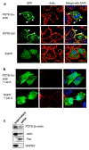
POTE-2α-actin and POTE-2γC are associated with actin protein in Hela cells. a Hela cells were transfected with pPOTE-2α-actin-EGFP, pPOTE-2γC-EGFP or pEGFP vector. After 24 h of transfection, cells were fixed with 4% PFA and stained with phalloidin-TRITC (red) and DAPI (blue). Yellow shows merged image of co-localization of endogenous actin and POTE-2α-actin-EGFP or POTE-2γC-EGFP. b POTE localization is dependent on intact actin filaments. Hela cells transfected with POTE-2α-actin-EGFP were treated Lat A for 30 min before PFA fixation. Cells were then stained with phalloidin-TRITC and DAPI for 5 min and analyzed by confocal microscopy. c Hela cells were lysed with hypotonic buffer as described in “Experimental procedures”. Equivalent volume of soluble and membrane fractions were analyzed by western blot with anti-POTE (PG5), anti-Fas (membrane marker), anti-actin or anti-GAPDH (soluble fraction marker) antibodies
Fig. 3
Fig. 3
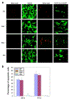
POTE over-expression induces apoptosis in Hela cells. a Hela cells transfected with pEGFP, pPOTE-2α-actin-EGFP or pPOTE-2γC-EGFP. At 2 or 3 days post-transfection, cells were incubated with RED-VAD-FMK for 1 h before fixation with 2% PFA. Cells were visualized with confocal microscopy. b Quantitative analysis of POTE induced apoptosis. Transfected cells were stained with RED-VAD-FMK for 1 h and red-staining cells and total transfected cells were counted under microscopy. Three separate experiments were repeated and each time about 100 transfected cells were counted
Fig. 4
Fig. 4
POTE induced apoptosis is dependent on pro-apoptotic protein Bak. a & b MEF cells, wild-type (wt), Bax−/−, Bak−/− and dko Bax−/− Bak−/−(double knock-out) were transfected with pEGFP or pPOTE-2γC-EGFP. At 2 or 3 days post-transfection, cells were incubated with RED-VAD-FMK for 1 h, and then fixed with 2% PFA for 3 min. Red labeled cells and total transfected cells were analyzed by confocal microscopy (a) or counted (b). Three separate experiments were performed and each time more than 300 cells were analyzed
Fig. 5
Fig. 5
Endogenous POTE-2α-actin expression is regulated by death receptor activation. a & b Hela cells at about 60% confluence were treated with anti-Fas antibody (a) or with 30 ng/ml of TRAIL (b) at the indicated concentrations and times before western blot analysis. Equal amount of cell lysate was analyzed by western blot. POTE-2α-actin was detected by PG5; GADPH as a loading control. c Hela cells were treated with 400 ng/ml anti-Fas antibody at the indicated times. RNA was prepared and real-time PCR was performed. The relative expression was quantified using actin RNA levels as the control

References

    1. Jin Z, El-Deiry SW (2005) Overview of cell death signaling pathways. Cancer Biol Ther 4:139–163 - PubMed
    1. O’ Brien DI, Nally K, Kelly RG, O’Connor TM, Shanahan F, O’Connell J (2005) Targeting the Fas/Fas ligand pathway in cancer. Expert Opin Ther Targets 9:1031–1044 - PubMed
    1. Nagata S, Golstein P (1995) The Fas death factor. Science 267:1449–1456 - PubMed
    1. Fletcher J, Huang D (2008) Controlling the cell death mediators Bax and Bak: puzzles and conundrums. Cell Cycle 7:39–44 - PubMed
    1. Reichmann E (2002) The biological role of the Fas/FasL system during tumor formation and progression. Semin Cancer Biol 12:309–315 - PubMed

Publication types

MeSH terms