. 2009 Dec;17(12):2096-102.
doi: 10.1038/mt.2009.181. Epub 2009 Aug 11.

Inner limiting membrane barriers to AAV-mediated retinal transduction from the vitreous

Affiliations

Inner limiting membrane barriers to AAV-mediated retinal transduction from the vitreous

Deniz Dalkara et al. Mol Ther. 2009 Dec.

Abstract

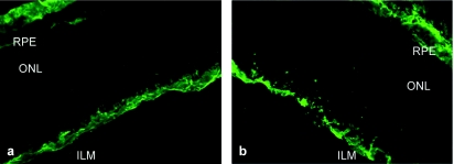
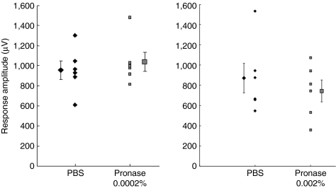
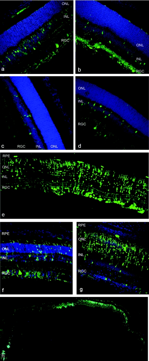
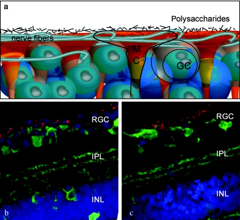
Adeno-associated viral gene therapy has shown great promise in treating retinal disorders, with three promising clinical trials in progress. Numerous adeno-associated virus (AAV) serotypes can infect various cells of the retina when administered subretinally, but the retinal detachment accompanying this injection induces changes that negatively impact the microenvironment and survival of retinal neurons. Intravitreal administration could circumvent this problem, but only AAV2 can infect retinal cells from the vitreous, and transduction is limited to the inner retina. We therefore sought to investigate and reduce barriers to transduction from the vitreous. We fluorescently labeled several AAV serotype capsids and followed their retinal distribution after intravitreal injection. AAV2, 8, and 9 accumulate at the vitreoretinal junction. AAV1 and 5 show no accumulation, indicating a lack of appropriate receptors at the inner limiting membrane (ILM). Importantly, mild digestion of the ILM with a nonspecific protease enabled substantially enhanced transduction of multiple retinal cell types from the vitreous, with AAV5 mediating particularly remarkable expression in all retinal layers. This protease treatment has no effect on retinal function as shown by electroretinogram (ERG) and visual cortex cell population responses. These findings may help avoid limitations, risks, and damage associated with subretinal injections currently necessary for clinical gene therapy.


Figures

<b>Figure 1</b>
Figure 1
Morphological effects of Pronase E on the inner limiting membrane (ILM). (a) Untreated retina stained with anti-laminin antibody shows immunoreactivity at the ILM and at the choroid, whereas (b) laminin immunolabeling after treatment with 0.01% Pronase E exhibits a disintegrated ILM structure. ONL, outer nuclear layer; RPE, retinal pigment epithelium.
<b>Figure 2</b>
Figure 2
The electroretinogram of animals injected with 0.0002% (n = 8), 0.001% (n = 6), and 0.002% (n = 8) Pronase E was analyzed to assess toxicity of mild enzymatic cleavage of the inner limiting membrane (ILM). Each animal was injected with the enzyme in the vitreous of one eye and phosphate-buffered saline (PBS) in the contralateral eye. Pronase E concentrations are (a) 0.0002%, (b) 0.001%, and (c) 0.002%. Pronase E injection exhibited no significant change in A- or B-wave amplitude compared to control PBS-injected eyes. Statistical differences between Pronase E and PBS-injected eyes were calculated by Student's t-test.
<b>Figure 3</b>
Figure 3
Peak amplitude of visually evoked potentials in response to full-field stimulation of eyes injected with phosphate-buffered saline (PBS) (diamonds) or Pronase (squares) at the low dose, n = 6, and high dose, n = 6. Recordings were performed on the contralateral visual cortex. Means for each data set are shown laterally displaced. Error bars indicate SEM. The two data sets for each dose were not significantly different (n = 6, P > 0.6, Wilcoxon signed-rank test).
<b>Figure 4</b>
Figure 4
Green fluorescent protein (GFP) expression in cryosections of rat retina after intravitreal delivery of 1011 vector genomes of adeno-associated virus (AAV) vectors carrying smCBA.hGFP in the presence of 0.0002% Pronase, 3 weeks after injection. Nuclei are stained with 4′,6-diamidino-2-phenylindole, shown in blue. (a) AAV1, (b) AAV2, (c) AAV8, (d) AAV9, and (e–h) AAV5. A representative area shows robust GFP fluorescence in all retinal layers in e. The proportion of transduced cells shows variability from one part to the other (n = 6) with strong expression in retinal ganglion cells (RGCs) and Müller cells in f and predominantly photoreceptors with weaker RGC with some expression in the RPE in g. An entire cryoslice is shown in h.
<b>Figure 5</b>
Figure 5
AAV particle localization at the vitreoretinal junction. (a) Schematic representation of the overlapping structures of the vitreoretinal junction. (b) Confocal images of AAV2-Cy3 and (c) AAV9-Cy3 accumulation at the vitreoretinal junction (red). The cryosections are counterstained with an antibody against calbindin (green), which labels the retinal neurons.

References

    1. Mueller C., and , Flotte TR. Clinical gene therapy using recombinant adeno-associated virus vectors. Gene Ther. 2008;15:858–863. - PubMed
    1. Lebherz C, Maguire A, Tang W, Bennett J., and , Wilson JM. Novel AAV serotypes for improved ocular gene transfer. J Gene Med. 2008;10:375–382. - PMC - PubMed
    1. Hauswirth WW, Aleman TS, Kaushal S, Cideciyan AV, Schwartz SB, Wang L, et al. Treatment of leber congenital amaurosis due to RPE65 mutations by ocular subretinal injection of adeno-associated virus gene vector: short-term results of a phase I trial. Hum Gene Ther. 2008;19:979–990. - PMC - PubMed
    1. Büning H, Perabo L, Coutelle O, Quadt-Humme S., and , Hallek M. Recent developments in adeno-associated virus vector technology. J Gene Med. 2008;10:717–733. - PubMed
    1. Goncalves MA. Adeno-associated virus: from defective virus to effective vector. Virol J. 2005;2:43. - PMC - PubMed

Publication types

LinkOut - more resources