

Prognostic value of Dicer expression in human breast cancers and association with the mesenchymal phenotype
- PMID: 19672267
- PMCID: PMC2736830
- DOI: 10.1038/sj.bjc.6605193
Prognostic value of Dicer expression in human breast cancers and association with the mesenchymal phenotype
Abstract
Background: Dicer, a ribonuclease, is the key enzyme required for the biogenesis of microRNAs and small interfering RNAs and is essential for both mammalian development and cell differentiation. Recent evidence indicates that Dicer may also be involved in tumourigenesis. However, no studies have examined the clinical significance of Dicer at both the RNA and the protein levels in breast cancer.
Methods: In this study, the biological and prognostic value of Dicer expression was assessed in breast cancer cell lines, breast cancer progression cellular models, and in two well-characterised sets of breast carcinoma samples obtained from patients with long-term follow-up using tissue microarrays and quantitative reverse transcription-PCR.
Results: We have found that Dicer protein expression is significantly associated with hormone receptor status and cancer subtype in breast tumours (ER P=0.008; PR P=0.019; cancer subtype P=0.023, luminal A P=0.0174). Dicer mRNA expression appeared to have an independent prognostic impact in metastatic disease (hazard ratio=3.36, P=0.0032). In the breast cancer cell lines, lower Dicer expression was found in cells harbouring a mesenchymal phenotype and in metastatic bone derivatives of a breast cancer cell line. These findings suggest that the downregulation of Dicer expression may be related to the metastatic spread of tumours.
Conclusion: Assessment of Dicer expression may facilitate prediction of distant metastases for patients suffering from breast cancer.
Figures
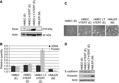
 ). Morphological characteristics and expression of epithelial and mesenchymal markers in the human breast tumour progression cellular model: (C) Morphological characteristics of the HMEC+hTERT, HMEC+LT+hTERT, and HMLER cells in vitro as assessed by phase-contrast microscopy. Images are shown at × 40 magnification. (D) Western blot analysis of E-cadherin and vimentin expression in the four human breast cell lines, HMEC, HMEC+hTERT, HMEC+LT+hTERT, and HMLER. Actin served as a loading control. E, epithelial phenotype; M, mesenchymal phenotype.
 ). E, epithelial phenotype; M mesenchymal phenotype; E/M, mixed phenotype. Expression of Dicer in two bone metastatic derivatives of MDA-MB-231 cells: (C) western blot analysis of Dicer expression in the three cell lines, namely MDA-MB-231, BO2, and SCP2. The signal intensity of Dicer was normalised to that of actin. (D) The relative levels of Dicer mRNA were measured by real-time RT–PCR, and each bar represents the mean±s.d. of the PCRs in triplicate (▪). Dicer protein levels were quantified using Quantity One software (BioRad, Marnes-la-Coquette, France) and expressed as protein relative quantity. The ratios of Dicer/actin of three independent studies were expressed as mean±s.d. (
 ).
).
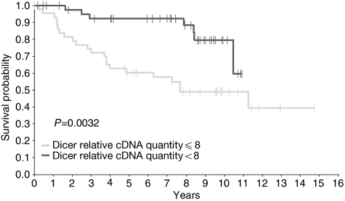
 >8 cDNA relative quantity (7 out of 43 progressions; 8-year event-free survival probability 88.3% (95% CI: 77.3–99.3);
  ⩽8 cDNA relative quantity (22 out of 45 progressions; 8-year event-free survival probability 49.1% (95% CI: 33.6–64.6); log-rank test: P=0.0032.
References
-
- American Cell Type Collection. http://www.ATCC.org
-
- Aslakson CJ, Miller FR (1992) Selective events in the metastatic process defined by analysis of the sequential dissemination of subpopulations of a mouse mammary tumor. Cancer Res 52: 1399–1405 - PubMed
-
- Bellahcène A, Bachelier R, Detry C, Lidereau R, Clézardin P, Castronovo V (2007) Transcriptome analysis reveals an osteoblast-like phenotype for human osteotropic breast cancer cells. Breast Cancer Res Treat 101: 135–148 - PubMed
-
- Bernstein E, Kim SY, Carmell MA, Murchison EP, Alcorn H, Li MZ, Mills AA, Elledge SJ, Anderson KV, Hannon GJ (2003) Dicer is essential for mouse development. Nat Genet 35: 215–217, Erratum in: Nat Genet. 2003 35:287 - PubMed
Publication types
MeSH terms
Substances
LinkOut - more resources
Full Text Sources
Other Literature Sources
Medical
Research Materials
