

Investigation of the feasibility of relative 3D dosimetry in the Radiologic Physics Center Head and Neck IMRT phantom using presage/optical-CT
- PMID: 19673232
- PMCID: PMC2832043
- DOI: 10.1118/1.3148534
Investigation of the feasibility of relative 3D dosimetry in the Radiologic Physics Center Head and Neck IMRT phantom using presage/optical-CT
Abstract
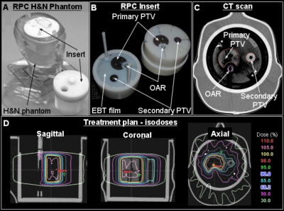
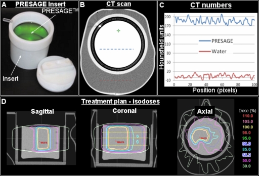
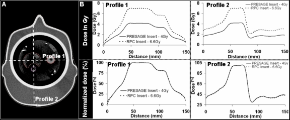
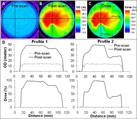
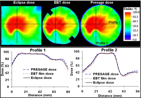
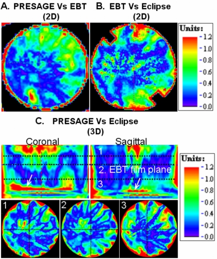
This study presents the application of the Presage/optical-CT 3D dosimetry system for relative dosimetry in the Radiologic Physics Center (RPC) Head and Neck (H&N) IMRT phantom. Performance of the system was evaluated by comparison with the "gold-standard" RPC credentialing test. A modified Presage cylindrical insert was created that extended the capability of the RPC H&N phantom to 3D dosimetry. The RPC phantom was taken through the entire treatment planning procedure with both the standard RPC insert and the modified Presage insert. An IMRT plan was created to match the desired dose constraints of the credentialing test. This plan was delivered twice to the RPC phantom: first containing the standard insert, and then again containing the Presage insert. After irradiation, the standard insert was sent for routine credentialing analysis; including point dose measurements (TLD) and planar Gafchromic EBT film measurement. The 3D dose distribution from Presage was read out at Duke using the OCTOPUS 5X optical-CT scanner. The Presage distribution was compared with gold-standard EBT measurement (determined by the RPC) and the calculated Eclipse distribution. The agreement between the normalized EBT, Presage, and Eclipse distributions, in the central axial plane was evaluated using profiles and gamma-map comparisons (4% dose difference and 3 mm distance to agreement). Profiles showed good agreement between EBT, Presage, and Eclipse distributions. 2D gamma-map comparisons between all three modalities showed at least 98% pass rate. The excellent agreement between Presage and EBT in the central plane established Presage as a standard against which to evaluate the accuracy of the 3D calculated Eclipse distribution. A gamma comparison between normalized Presage and Eclipse 3D distributions gave an overall pass rate of approximately 94%. In conclusion, the Presage/optical-CT system was found to be feasible for relative 3D dosimetry in the RPC IMRT H&N phantom. The potential to extend the RPC IMRT credentialing procedure to 3D may be feasible provided accurate calibration to dose (Gy) and robustness to shipping stress are demonstrated.
Figures
References
-
- Molineu A. et al. , “Design and implementation of an anthropomorphic quality assurance phantom for intensity-modulated radiation therapy for the Radiation Therapy Oncology Group,” Int. J. Radiat. Oncol., Biol., Phys. 63, 577–583 (2005). - PubMed
-
- Babic S., Battista J., and Jordan K., “Three-dimensional dose verification for intensity-modulated radiation therapy in the radiological physics centre head-and-neck phantom using optical computed tomography scans of ferrous xylenol-orange gel dosimeters,” Int. J. Radiat. Oncol., Biol., Phys. 70, 1281–1291 (2008). - PubMed
Publication types
MeSH terms
Grants and funding
LinkOut - more resources
Full Text Sources
Medical
