Review
. 2009 Nov;11(11):1561-70.
doi: 10.1111/j.1462-5822.2009.01363.x. Epub 2009 Aug 5.

Morphogenesis of hepatitis B virus and its subviral envelope particles

Affiliations
Review

Morphogenesis of hepatitis B virus and its subviral envelope particles

Romuald Patient et al. Cell Microbiol. 2009 Nov.

Abstract

After cell hijacking and intracellular amplification, non-lytic enveloped viruses are usually released from the infected cell by budding across internal membranes or through the plasma membrane. The enveloped human hepatitis B virus (HBV) is an example of virus using an intracellular compartment to form new virions. Four decades after its discovery, HBV is still the primary cause of death by cancer due to a viral infection worldwide. Despite numerous studies on HBV genome replication little is known about its morphogenesis process. In addition to viral neogenesis, the HBV envelope proteins have the capability without any other viral component to form empty subviral envelope particles (SVPs), which are secreted into the blood of infected patients. A better knowledge of this process may be critical for future antiviral strategies. Previous studies have speculated that the morphogenesis of HBV and its SVPs occur through the same mechanisms. However, recent data clearly suggest that two different processes, including constitutive Golgi pathway or cellular machinery that generates internal vesicles of multivesicular bodies (MVB), independently form these two viral entities.


Figures
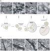
FIG. 1
FIG. 1
EM observation of BHK-21 cells producing the HBV S protein (yellow dot, materials and methods described in Patient et al, 2007) and model for the SVP morphogenesis and traffic. Vesicles about 0.2 to 0.3 μm in diameter (arrows), packed with 20 nm-large HBV S filaments, are observed budding from the nuclear envelope (A-a). After budding, these vesicles are packed with HBV S filaments and appear in lengthwise or crosswise sections with a crystal-like structure (A-b). The fusion of such vesicles with the endoplasmic reticulum-Golgi intermediate compartment (ERGIC) provides the relaxing of these filaments from their crystal-like structure in the ERGIC lumen (A-c). Negative staining of the purified intracellular HBV S filaments shows that these filaments tended to dissociate at their ends (arrow) into subviral spherical particles (A-d). A careful observation of the cell perinuclear space suggestes that nascent filaments are self-assembled in the lumen of this compartment rather than by membrane-derived budding (B-a to B-d). For some filaments, intraluminal free ends are identified (arrow in B-d). Bars in A-a, A-b, A-c, B-a, B-b and B-c, 200 nm; Bars in A-d and B-d, 100 nm; Nu, nucleus; Cy, cytosol; the cellular chaperones potentially involved in the SVP morphogenesis are presented as black zig-zag.
FIG. 2
FIG. 2
Model for HBV morphogenesis. On the left is the representation of a typical endocytosis and multivesicular bodies (MVB) sorting of an activated membrane protein, e.g. the Epithelium Growth Factor Receptor (EGFR). The ubiquitylated (Ub, black square) protein is transported into an early endosome to the ESCRT machinery. In MVB, the ubiquitylated receptor is recognized by ESCRT-I (I, blue) which immediately recruits additional components of the MVB pathway: ESCRT-II (II, red), ESCRT-III (III, green) which is anchored to the MVB membrane by myristylation (zig-zag), Alix/AIP1 (Alix, purple) and Vps4A/B (brown) which is critical for disassembly of the complex following inward budding of intraluminal vesicle (IVL) into the MVB lumen. On the right, the hijacking of MVB sorting machinery for HBV morphogenesis is depicted. See text for details. pgRNA, pre-genomic RNA; gDNA, genomic DNA; the viral reverse-transcriptase located inside the capsid is presented as a black heptagon.

References

    1. Appenzeller-Herzog C, Hauri HP. The ER-Golgi intermediate compartment (ERGIC): in search of its identity and function. J Cell Sci. 2006;119:2173–2183. - PubMed
    1. Beck J, Nassal M. Hepatitis B virus replication. World J Gastroenterol. 2007;13:48–64. - PMC - PubMed
    1. Berting A, Hahnen J, Kroger M, Gerlich WH. Computer-aided studies on the spatial structure of the small hepatitis B surface protein. Intervirology. 1995;38:8–15. - PubMed
    1. Blumberg BS, Alter HJ, Visnich S. A “New” Antigen in Leukemia Sera. Jama. 1965;191:541–546. - PubMed
    1. Bruss V. Hepatitis B virus morphogenesis. World J Gastroenterol. 2007;13:65–73. - PMC - PubMed