

Surface area congruence of atlas superior articulating facets and occipital condyles
- PMID: 19674714
- PMCID: PMC2647103
- DOI: 10.1016/j.jcme.2007.08.007
Surface area congruence of atlas superior articulating facets and occipital condyles
Abstract
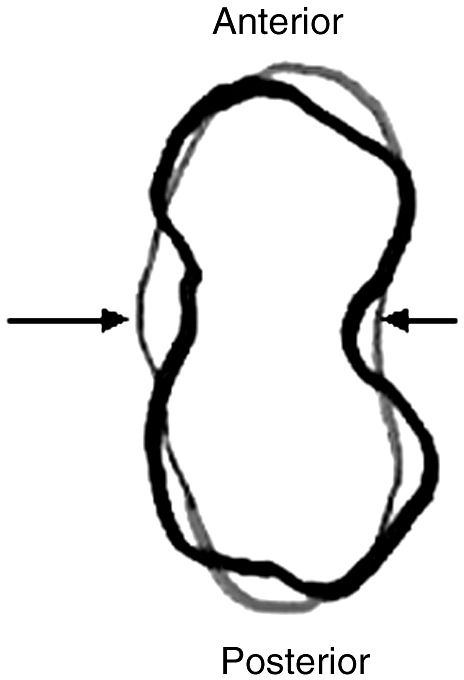
Objective: The purpose of this study was to compare the surface areas of 12 atlanto-occipital joints from 6 cadavers to determine how well their ipsilateral and contralateral surface areas matched.
Methods: Three methods were used. Method 1 consisted of digitized photographs downloaded to the Able Image Analyser software program (Ljubljana, Slovenia; http://able.mulabs.com). The perimeters were measured and expressed as square millimeters. Method 2 consisted of a point count method using moulds of the joint surfaces produced by pressing aluminum paper, leaving a clear imprint for analysis. Method 3 consisted of drawing outlines of the molds from method 2 onto transparencies and assessing overlap.
Results: Method 1 showed a moderate correlation of matched articular surfaces between the left C1 superior articulating surface and the left condyle (r = 0.573, P = .01). Method 2 showed moderate correlations between all surface areas that were analyzed. The matched pairs compared were left C1 superior articulating surface and left condyle (r = 0.588, P = .01) and right C1 and right condyle (r = 0.730, P = .001). The contralateral surfaces correlated were left C1 and right C1 (r = 0.596, P = .009) and right condyle and left condyle (r = 0.769, P = .000). Method 3 showed no statistically significant differences between surface areas.
Conclusions: All 3 methods revealed that the articular surfaces of the atlas and corresponding or contralateral condyle for specimens used in this study were not an exact match.
Figures

References
-
- Addington E.A. Overview of Blair cervical technique. Prepared for the Council on Chiropractic Practice. Chandler, Arizona. http://www.chiro.org/LINKS/blair.html October 1995 [cited 2005 Aug 5]. Available from:
-
- Addington E.A.Blair cervical spinographic analysisProceedings from the Conference on Research and Education; 1991 June: Monterey, CA. Foundation for Chiropractic Education and Research1991
-
- Remier P.A. Modern X-ray practice and spinography. The Palmer School of Chiropractic; Davenport (Iowa): 1957. pp. 312–378.
-
- Addington E.A. Blair Chiropractic Society. The Blair technique [homepage on the Internet]. X-rays explained. http://blairchiropractic.com/xrays_explained.htm June 1991[cited 2007 Jun 18] Available from:
-
- Blair W.G. Research; for evaluation; for progress. Intl Rev Chiropr. 1968;22(8):8–11.
LinkOut - more resources
Full Text Sources
