

New approach for m-cell-specific molecules screening by comprehensive transcriptome analysis
- PMID: 19675110
- PMCID: PMC2725790
- DOI: 10.1093/dnares/dsp013
New approach for m-cell-specific molecules screening by comprehensive transcriptome analysis
Abstract
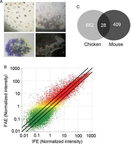
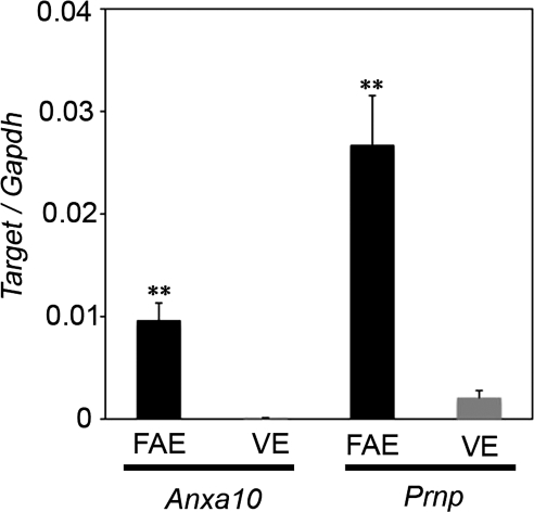
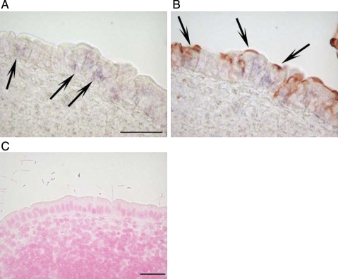
A minor population of M cells within the follicle-associated epithelium (FAE) of intestinal Peyer's patches (PPs) serves as a major portal for entry of exogenous antigens. Characterization of the mammalian M cells, including identification of M-cell surface molecules used for bacterial uptake, has been hampered by their relative rarity. In contrast, M cells constitute virtually all of the FAE cells in the avian bursa of Fabricius. We therefore performed comparative gene expression profiling of chicken and murine FAE to identify commonly expressed genes by M cells in both species. The comprehensive transcriptome analysis revealed that 28 genes were commonly up-regulated in FAE from both species. In situ hybridization revealed that annexin A10 (Anxa10) mRNA was scattered in FAE, and co-localized with Ulex europaeus agglutinin-1 binding to M cells. Whole-mount immunostaining also revealed that cellular prion protein (PrP(C)) was expressed on the luminal side of the apical plasma membrane of M cells, and co-localized with grycoprotein 2 that recognizes only M cells in murine PP. Our findings identify new M-cell-specific molecules through using comprehensive transcriptome analysis. These conserved molecules in M cells of mice and chickens may play essential roles in M-cell function and/or differentiation.
Figures


Similar articles
-
Oral Prion Neuroinvasion Occurs Independently of PrPC Expression in the Gut Epithelium.J Virol. 2018 Sep 12;92(19):e01010-18. doi: 10.1128/JVI.01010-18. Print 2018 Oct 1. J Virol. 2018. PMID: 30021891 Free PMC article.
-
Peptidoglycan recognition protein expression in mouse Peyer's Patch follicle associated epithelium suggests functional specialization.Cell Immunol. 2003 Jul;224(1):8-16. doi: 10.1016/s0008-8749(03)00155-2. Cell Immunol. 2003. PMID: 14572796
-
Distinct gene expression profiles characterize cellular phenotypes of follicle-associated epithelium and M cells.DNA Res. 2005;12(2):127-37. doi: 10.1093/dnares/12.2.127. DNA Res. 2005. PMID: 16303744
-
Comprehensive gene expression profiling of Peyer's patch M cells, villous M-like cells, and intestinal epithelial cells.J Immunol. 2008 Jun 15;180(12):7840-6. doi: 10.4049/jimmunol.180.12.7840. J Immunol. 2008. PMID: 18523247
-
Development of Peyer's patches, follicle-associated epithelium and M cell: lessons from immunodeficient and knockout mice.Semin Immunol. 1999 Jun;11(3):183-91. doi: 10.1006/smim.1999.0174. Semin Immunol. 1999. PMID: 10381864 Review.
Cited by
-
Oral Prion Neuroinvasion Occurs Independently of PrPC Expression in the Gut Epithelium.J Virol. 2018 Sep 12;92(19):e01010-18. doi: 10.1128/JVI.01010-18. Print 2018 Oct 1. J Virol. 2018. PMID: 30021891 Free PMC article.
-
Prion pathogenesis and secondary lymphoid organs (SLO): tracking the SLO spread of prions to the brain.Prion. 2012 Sep-Oct;6(4):322-33. doi: 10.4161/pri.20676. Epub 2012 Aug 16. Prion. 2012. PMID: 22895090 Free PMC article.
-
How do PrPSc Prions Spread between Host Species, and within Hosts?Pathogens. 2017 Nov 24;6(4):60. doi: 10.3390/pathogens6040060. Pathogens. 2017. PMID: 29186791 Free PMC article. Review.
-
Establishing Boundaries: The Relationship That Exists between Intestinal Epithelial Cells and Gut-Dwelling Bacteria.Microorganisms. 2019 Dec 9;7(12):663. doi: 10.3390/microorganisms7120663. Microorganisms. 2019. PMID: 31818022 Free PMC article. Review.
-
Homeostatic mini-intestines through scaffold-guided organoid morphogenesis.Nature. 2020 Sep;585(7826):574-578. doi: 10.1038/s41586-020-2724-8. Epub 2020 Sep 16. Nature. 2020. PMID: 32939089
References
-
- Neutra M.R., Mantis N.J., Kraehenbuhl J.P. Collaboration of epithelial cells with organized mucosal lymphoid tissues. Nat. Immunol. 2001;2:1004–9. - PubMed
-
- Bockman D.E., Cooper M.D. Pinocytosis by epithelium associated with lymphoid follicles in the bursa of Fabricius, appendix, and Peyer's patches. An electron microscopic study. Am. J. Anat. 1973;136:455–77. - PubMed
-
- Owen R.L. Sequential uptake of horseradish peroxidase by lymphoid follicle epithelium of Peyer's patches in the normal unobstructed mouse intestine: an ultrastructural study. Gastroenterology. 1977;72:440–51. - PubMed
Publication types
MeSH terms
Substances
Associated data
- Actions
LinkOut - more resources
Full Text Sources
Molecular Biology Databases
Research Materials
Miscellaneous
