. 2009 Sep;136(18):3043-55.
doi: 10.1242/dev.039008. Epub 2009 Aug 12.

Systematic analysis of dynamic miRNA-target interactions during C. elegans development

Affiliations

Systematic analysis of dynamic miRNA-target interactions during C. elegans development

Liang Zhang et al. Development. 2009 Sep.

Abstract

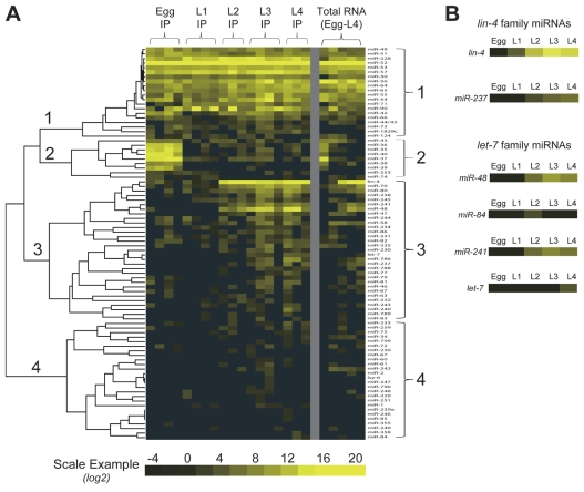
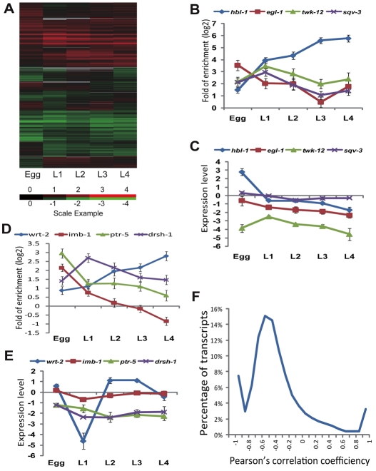
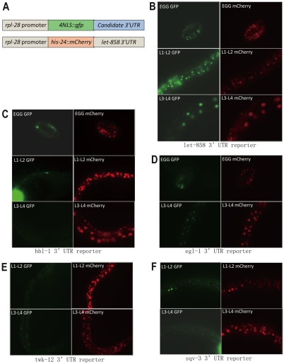
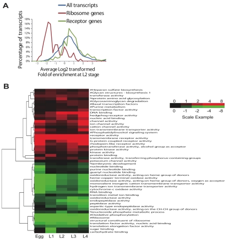
Although microRNA (miRNA)-mediated functions have been implicated in many aspects of animal development, the majority of miRNA::mRNA regulatory interactions remain to be characterized experimentally. We used an AIN/GW182 protein immunoprecipitation approach to systematically analyze miRNA::mRNA interactions during C. elegans development. We characterized the composition of miRNAs in functional miRNA-induced silencing complexes (miRISCs) at each developmental stage and identified three sets of miRNAs with distinct stage-specificity of function. We then identified thousands of miRNA targets in each developmental stage, including a significant portion that is subject to differential miRNA regulation during development. By identifying thousands of miRNA family-mRNA pairs with temporally correlated patterns of AIN-2 association, we gained valuable information on the principles of physiological miRNA::target recognition and predicted 1589 high-confidence miRNA family::mRNA interactions. Our data support the idea that miRNAs preferentially target genes involved in signaling processes and avoid genes with housekeeping functions, and that miRNAs orchestrate temporal developmental programs by coordinately targeting or avoiding genes involved in particular biological functions.


Figures

Fig. 1.
Fig. 1.
Dynamic association of miRNAs with AIN-2-miRISCs during development. (A) Heat map illustration of log2-transformed miRNA concentrations measured by pyrosequencing in 18 AIN-2::GFP IP and five total RNA samples from worms at five indicated developmental stages. Each row represents a miRNA and each column represents an individual sample. Only the 86 detected miRNAs were included. Different columns for the same stage are different replicates for that stage. The heat map and tree structure were generated by the self-organization map approach followed by complete linkage hierarchical clustering using the cluster software. The miRNAs can be grouped into four major clusters: all-stage miRNAs (1), early-stage miRNAs (2), late-stage miRNAs (3) and miRNAs with sporadic low level expressions (4). (B) The expression pattern of lin-4 and let-7 family miRNAs determined by pyrosequencing. The results were taken directly from the total RNA samples displayed in A.
Fig. 2.
Fig. 2.
Dynamic pattern of fold of enrichments of miRNA targets in AIN-2 IP during development. (A) Heat map illustration of enrichment of the 15,013 testable transcripts in the AIN-2::GFP IP from worms at the five developmental stages. Log2-transformed stage-averaged fold of enrichment was displayed. Each row represents a transcript. Each column represents a stage as indicated. Red indicates enrichment in IP, green indicates depletion from IP, and gray indicates missing values. (B,D) Eight examples of dynamic miRNA::target interactions during development. The vertical axis represents log2-transformed stage-averaged fold of enrichment. (C,E) The mRNA expression pattern of the example genes in B,D during development. The vertical axis represent log2 transformed stage-averaged mRNA expression level, measured by dividing the microarray signal of the mRNA in each total lysate sample by the average signal of all mRNAs in the same sample. Error bars in B-E indicate s.e.m. (F) Distribution of the Pearson's correlation coefficiency between the expression level and the fold of enrichment in AIN-2 IP of all the testable transcripts during development. The vertical axis represents the percentage of transcripts within each 0.1 interval of the correlation coefficiency value. The median Pearson's correlation coefficiency value of all the testable transcripts is -0.46.
Fig. 3.
Fig. 3.
Reporter analysis of dynamics of miRNA regulation on the expression of four genes during development. (A) Cartoon illustration of the dual-color reporter analysis strategy. As the control for transgene expression, rpl-28 driving his-24::mCherry:let-858 3UTR was co-expressed with the GFP reporters in each animal from the same extra-chromosomal array. (B-F) Fluorescence images of living animals expressing GFP reporters of four indicated 3′UTRs. The stage of each image is indicated. All GFP reporters contain a 4× SV40 nuclear localization signal (NLS) and were driven by the ubiquitous rpl-28 promoter. The mCherry images were taken immediately after the GFP images of the same animals using exactly the same scope settings.
Fig. 4.
Fig. 4.
miRNA regulation prominently prefers genes involved in cell signaling and avoids genes of housekeeping functions during development. (A) Distribution of miRNA regulation intensity on selected gene classes at the L2 stage. The vertical axis represents the percentage of transcripts within each 0.25 interval of log2-transformed stage-averaged fold enrichment. Receptor genes represent genes annotated with GO term: receptor activity. Ribosome genes represent genes annotated with GO term: structural constituent of ribosome. (B) Heat map and hierarchical clustering display of absolute value of log10-transformed P-values for the association between biological labels and the relative miRNA regulation intensity based on Fatiscan analysis (see Table S3 in the supplementary material). Each column represents a stage as indicated. Each row represents a biological label as indicated. `#' indicates KEGG pathway annotations (Kanehisa, 1997; Kanehisa et al., 2006); `%' and all the other labels indicate GO: Biological Process annotations and GO: Molecular Function annotations, respectively (Ashburner et al., 2000). Only labels that were significantly preferred (red) or avoided (green; FDR adjusted, P<0.01) in at least one stage were displayed.
Fig. 5.
Fig. 5.
Matching AIN-2 association patterns of miRNA and mRNA during development greatly facilitates the search for specific miRNA::target interactions. (A) Fold of enrichment for different types of seed matches in the pattern-matched miRNA family::mRNA pairs containing annotated 3′UTRs compared with all other testable miRNA family::mRNA pairs containing annotated 3′UTRs. Perfect 7-mer refers to a stretch of continuous 7-nucleotide that are a perfect complementary match to the 5′ end of the miRNA starting from position 2. Position 1 or position 3 7-mer means the 7-nucleotide perfect match started from position 1 or position 3 instead. 1 G:U 7-mer means there is one G:U wobble pair within the position 2-8 7-mer match. (B) The fold of enrichment for different types of seed matches in conserved potential miRNA family::mRNA-binding sites that are pattern matched versus those that are not pattern matched. (C) The fold of enrichment for different total interaction energy in conserved potential miRNA family::mRNA-binding sites that are pattern matched versus those that are not pattern matched. (D) The fold of enrichment for different total interaction energy in conserved potential miRNA family::mRNA-binding sites with particular seed configurations that are pattern matched versus those that are not pattern matched. 7-mer refers to any miRNA::target helix that has perfect position 2-8 complementarity, which included the position 2-8 7-mers, position 2-9 8-mers, and position 1-8 8-mers from A. `Non 7-mers' includes all other seed types in A. (E,F) Fluorescence images of live animals expressing the GFP reporter of the egl-1 3′UTR in a wild-type (E) or a mir-35 family overexpression (mir-35 ++, F) background. The stage of each pictured animal is indicated. See Fig. 3 for information regarding the reporter system.

References

    1. Abbott, A. L., Alvarez-Saavedra, E., Miska, E. A., Lau, N. C., Bartel, D. P., Horvitz, H. R. and Ambros, V. (2005). The let-7 MicroRNA family members mir-48, mir-84, and mir-241 function together to regulate developmental timing in Caenorhabditis elegans. Dev. Cell 9, 403-414. - PMC - PubMed
    1. Abrahante, J. E., Daul, A. L., Li, M., Volk, M. L., Tennessen, J. M., Miller, E. A. and Rougvie, A. E. (2003). The Caenorhabditis elegans hunchback-like gene lin-57/hbl-1 controls developmental time and is regulated by microRNAs. Dev. Cell 4, 625-637. - PubMed
    1. Al-Shahrour, F., Diaz-Uriarte, R. and Dopazo, J. (2005a). Discovering molecular functions significantly related to phenotypes by combining gene expression data and biological information. Bioinformatics 21, 2988-2993. - PubMed
    1. Al-Shahrour, F., Minguez, P., Vaquerizas, J. M., Conde, L. and Dopazo, J. (2005b). BABELOMICS: a suite of web tools for functional annotation and analysis of groups of genes in high-throughput experiments. Nucleic Acids Res. 33, W460-W464. - PMC - PubMed
    1. Al-Shahrour, F., Minguez, P., Tarraga, J., Montaner, D., Alloza, E., Vaquerizas, J. M., Conde, L., Blaschke, C., Vera, J. and Dopazo, J. (2006). BABELOMICS: a systems biology perspective in the functional annotation of genome-scale experiments. Nucleic Acids Res. 34, W472-W476. - PMC - PubMed

Publication types

MeSH terms