. 2009 Sep;136(18):3121-9.
doi: 10.1242/dev.033597. Epub 2009 Aug 12.

Receptor tyrosine phosphatases control tracheal tube geometries through negative regulation of Egfr signaling

Affiliations

Receptor tyrosine phosphatases control tracheal tube geometries through negative regulation of Egfr signaling

Mili Jeon et al. Development. 2009 Sep.

Abstract

The formation of epithelial tubes with defined shapes and sizes is essential for organ development. We describe a unique tracheal tubulogenesis phenotype caused by loss of both Drosophila type III receptor tyrosine phosphatases (RPTPs), Ptp4E and Ptp10D. Ptp4E is the only widely expressed Drosophila RPTP, and is the last of the six fly RPTPs to be genetically characterized. We recently isolated mutations in Ptp4E, and discovered that, although Ptp4E null mutants have no detectable phenotypes, double mutants lacking both Ptp4E and Ptp10D display synthetic lethality at hatching owing to respiratory failure. In these double mutants, unicellular and terminal tracheal branches develop large bubble-like cysts that selectively incorporate apical cell surface markers. Cysts in unicellular branches are enlargements of the lumen that are sealed by adherens junctions, whereas cysts in terminal branches are cytoplasmic vacuoles. Cyst size and number are increased by tracheal expression of activated Egfr tyrosine kinase, and decreased by reducing Egfr levels. Ptp10D forms a complex with Egfr in transfected cells. Downregulation of Egfr signaling by the RPTPs is required for the construction of tubular lumens, whether extracellular or intracellular, by cells that undergo remodeling during branch morphogenesis. The Ptp4E Ptp10D phenotype represents the first evidence of an essential role for RPTPs in epithelial organ development. These findings might be relevant to organ development and disease in mammals, because PTPRJ (DEP-1), an ortholog of Ptp4E/Ptp10D, interacts with the hepatocyte growth factor receptor tyrosine kinase. PTPRJ corresponds to the murine Scc1 (suppressor of colon cancer) gene.


Figures

Fig. 1.
Fig. 1.
Tracheal cyst phenotype in Ptp4E Ptp10D mutant embryos, and apical localization of Ptp10D and Ptp4E in tracheae. (A,B) Stage 15 Ptp4E1 Ptp10D1 (A) and wild-type (WT) (B) whole-mount embryos stained with the tracheal lumen marker antibody mAb 2A12, using HRP immunohistochemistry for visualization. Note that Fig. 6 shows that the 2A12 antigen (visualized by immunofluorescence) is reduced in Ptp4E Ptp10D unicellular tracheal branches relative to in wild type; however, we were still able to use 2A12 to visualize these branches using HRP immunohistochemistry. The HRP reaction can be carried out until saturation is reached, and under these conditions quantitative differences will not be detected; low and high-level expression will have the same appearance. (A) VBs and TC/LT branch junction cysts are indicated. Smaller cysts are found along LT branches and GBs. The middle section of the DT is convoluted. (C-E) Localization of Ptp10D and Ptp4E. (C) In stage 15 embryos, anti-Ptp10D mAb 8B2 labels the tracheae and the CNS axon ladder (arrow). (D,E) DT segments at stage 15. (D) Ptp10D (magenta) overlaps with the apical surface marker Sas (green). (E) UAS-Ptp4E-GFP (green), expressed in the trachea from Btl-Gal4, localizes to the apical membrane and overlaps with Ecad (magenta), which is apical in the DT. There are also bright Ptp4E-GFP spots on peripheral membranes (arrows). D and E are single confocal sections. Scale bars: in A, 40 μm for A,B; in C, 50 μm; in D,E, 5 μm.
Fig. 2.
Fig. 2.
Cysts are found only in unicellular and intracellular tubes. (A) Schematic representation of a tracheal metamer at stage 15. The branch names are labeled and color coded. (B) Cross-sections of tracheal tube types are diagrammed. Multicellular tubes consist of two or more cells surrounding the lumen. Unicellular tubes consist of a single cell wrapped around the lumen that forms autocellular junctions with itself. Intracellular tubes are composed of lumen within the cytoplasm; they lack cell junctions. DT and the proximal portion of TC are multicellular tubes, LG is an intracellular tube, and all other branches primarily consist of unicellular tubes with autocellular AJs. (C) Wild-type and Ptp4E1 Ptp10D1 dissected embryo fillets at stage 14, stained for the apical marker Crb. Two hemisegments are shown, with anterior to the left and dorsal up. In the mutant, the TC/LT branch junctions (asterisks) and the extending anterior branch of LT (arrow) are enlarged. (D) Wild-type and Ptp4E1 Ptp10D1 fillets at stage 15, stained for Crb. TC/LT branch junctions (asterisks) in the mutant have large cysts. GBs have 4-5 cysts per branch (arrows). (E) Cysts are found in LG terminal branches in the mutant. Two hemisegments are shown for wild-type and Ptp4E1 Ptp10D1 embryos, stained for the apical surface marker Sas. LGs are circled. Scale bars in C-E: 20 μm.
Fig. 3.
Fig. 3.
Rescue of the phenotype by tracheal expression of Ptp4E or Ptp10D, and quantitation of cyst phenotypes in different genotypes. (A) Stage 15 embryo fillets were stained with anti-Sas to label the tracheae and imaged using confocal microscopy. Tr 4-7 were scored in stage 15 embryos. Measurements were made for the diameters of TC/LT branch junctions (orange arrow) and GBs (purple arrows). In control embryos (Ptp4EKG2328 Ptp10D1/+), two measurements were made per GB, while in mutants all swellings on the branch were measured. (B-D) Expression of wild-type Ptp4E-GFP or Ptp10D in Ptp4E Ptp10D embryos fully rescued the cyst phenotype, as shown by all three bar graphs. There are no GB cysts at all in rescued or control embryos, so no bars are shown in the top three rows of C. All double mutant genotypes displayed similar phenotypes when GB cyst number and GB diameter metrics were evaluated. When TC/LT branch junction diameters were compared, Ptp4E1Ptp10D1 and Df(1)ovo4 Df(1)Δ59 showed larger measurements than the others. See Table S1 in the supplementary material for numerical data. The rescuing Ptp4E-GFP protein was expressed in tracheae and visualized in the experiment of Fig. 1. Ptp4E and Ptp10D alleles: Ptp4E1, N-terminal coding sequences deleted; Ptp4E (KG2328), P-element insertion; Df(1) ovo4, smallest deletion removing Ptp4E; Ptp10D1, N-terminal coding sequences deleted; Ptp10D (EP1172), P-element insertion; Df(1) Δ59, smallest deletion, removes Ptp10D and neighboring bif.
Fig. 4.
Fig. 4.
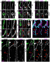
Apical membrane markers accumulate in cysts. (A-F) GBs were labeled for apical markers Crb (A-F) and Sas (A,B), basolateral marker Nrx (C,D), and junctional marker Ecad (E,F). In wild type (A,C,E), apical and junctional markers label a line, and Nrx labels a thin line and the junction between adjacent cells in the branch (C, arrow). In Ptp4E1 Ptp10D1, Crb and Sas are sequestered into the cyst (B), but Nrx is not (D). Ecad labels a line running through the cyst, indicating that an autocellular AJ is present; there is also weak labeling in the cyst that overlaps with Crb (F, arrow). (G-J) Membrane GFP (mGFP) was expressed in the tracheae and visualized with anti-GFP (green), together with anti-Crb (magenta) to label cysts. (G,H) Three cells in the GB are shown at high magnification (asterisks mark positions of nuclei). In wild-type (G), Crb localizes to the apical membrane and outlines the lumen, thus appearing as a line. In Ptp4E1 Ptp10D1 (H), a Crb-labeled cyst appears to be enclosed within the mGFP-labeled cell boundary (arrow). (I,J) High magnification views of three terminal cells (asterisks indicate nuclei), each of which forms an LG terminal branch, are shown. In wild type (I), lines indicate three cytoplasmic LG tubes. In Ptp4E1 Ptp10D1 (J), Crb labels cysts (arrows) located within the cell cytoplasm; the Crb lumen lines are missing in cells displaying cysts. (K) A Ptp4E1 Ptp10D1 stage 14 embryo expressing nuclear GFP (nGFP) in the tracheae was stained with anti-GFP (blue) and anti-Crb (magenta) antibodies. Cysts on GBs, indicated with arrows, are located adjacent to nuclei. At TC/LT branch junctions, cysts are surrounded by multiple nuclei (asterisks). (L,M) The TC/LT branch junction (asterisks) and the GB of a single hemisegment are shown in wild type (L) and Ptp4E1 Ptp10D1 (M), labeled for Crb (green) and Ecad (magenta). In the mutant, the TC/LT branch junction (M, asterisks) has multiple Ecad lines. Crb-labeled cysts in LT branches and GBs have normal Ecad lines running through them (M, arrows). Ecad also labels a ring-like structure (M, line) at the end of the GB. All panels are confocal z-stack projections. Scale bars: 5 μm in A-J; 20 μm in K; 50 μm in L,M.
Fig. 5.
Fig. 5.
TEM analysis shows that cysts are dilated lumens. Ultrastructural analysis of LT branch cross-sections in horizontal thin sections of stage 15 embryos was performed using TEM. (A) A wild-type tracheal branch with a single autocellular junction, and (D) a higher magnification view of the lumen and junction are shown. The AJ appears as a double electron-dense band between cell boundaries. The lumen contains diffuse electron-dense material. (B,C,E,F) Ptp4E1 Ptp10D1 tracheal branch cross-sections with a single autocellular junction and one nucleus (B,E), and with two cell junctions and two nuclei (C,F) are shown. The cyst in C,F is presumably at a TC/LT branch junction. E and F are higher magnification images of the lumen and junction areas in B and C, respectively. The lumens in mutant cells lack electron-dense material. Scale bars: 0.5 μm in A; 1 μm in B; 2 μm in C; 0.2 μm in D; 0.2 μm in E; 0.2 μm in F. n, nucleus; L, lumen; ER, rough endoplasmic reticulum; m, mitochondria; AJ, adherens junction; ne, nuclear envelope; b, basal membrane; ap, apical membrane.
Fig. 6.
Fig. 6.
The luminal 2A12 antigen fails to accumulate in Ptp4E Ptp10D tracheal branches that develop cysts. Stage 15 embryos were labeled for Sas (green) and the luminal marker 2A12 (magenta). (A) In wild-type control embryos, 2A12 accumulates in the lumens of all branches that are labeled with Sas. (B) In double mutant embryos, the 2A12 signal is absent or reduced in lumens of unicellular branches that develop cysts (arrow), but is present at wild-type or near wild-type levels in the multicellular DT and proximal TC branches.
Fig. 7.
Fig. 7.
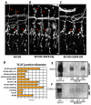
Enhancement and suppression of the Ptp4E Ptp10D phenotype by Egfr constructs, and physical association between Ptp10D and Egfr. (A-C) Three hemisegments of stage 15 embryos stained for Sas are shown for (A) Ptp4E1 Ptp10D1, (B) Ptp4E1Ptp10D1; Btl-Gal4::UAS-EgfrElp, and (C) Ptp4E1Ptp10D1; Btl-Gal4::UAS-Egfr-DN. (B) Constitutively activated EgfrElp expression in the tracheae causes dramatic enlargement of TC/LT branch junction cysts (asterisks). Almost all GB cells have cysts, and DBs, which have few cysts in unmodified mutants (<1 cyst/embryo) have many cysts (arrows) in the modified mutants. (C) Expression of dominant-negative Egfr partially suppresses the Ptp4E1 Ptp10D1 cyst phenotype. GBs show fewer cysts and TC/LT branch junction cysts are smaller than in Ptp4E1 Ptp10D1. (D) Quantitation of the phenotypes. TC/LT branch junction diameters are increased relative to those of Ptp4E1 Ptp10D1 alone when EgfrElp, wt Egfr, activated Btl, or activated Raf (Phl) are expressed; they are decreased when Egfr signaling is reduced by using Egfr-DN or Egfr-RNAi, or by crossing in one copy of an Egfr or a btl mutation. Expression of EgfrElp in a wild-type background does not produce enlargement of the TC/LT branch junctions relative to wild-type controls. (E,F) Complex formation between Ptp10D and Egfr was detected by coimmunoprecipitation from lysates of copper-induced S2 cells stably expressing Egfr and transiently expressing wild-type or substrate-trapping mutant Ptp10D (both driven from the MT promoter). (E) Immunoblotting with anti-Egfr shows the 180 kDa Egfr band when anti-Egfr or anti-Ptp10D was used for immunoprecipitation from cells expressing wild-type or mutant (DA) Ptp10D. (F) Immunoblotting with anti-Ptp10D shows a doublet when anti-Ptp10D or anti-Egfr was used for immunoprecipitation from cells expressing wild-type or mutant (DA) Ptp10D. The upper band of the doublet is ∼220 kDa, corresponding to the expected size of a glycosylated version of full-length Ptp10D, which is 1931 amino acids in length; these bands are not present in lysates from Ptp10D mutants (see Fig. S4 in the supplementary material).

Similar articles

Cited by

References

    1. Affolter, M. and Caussinus, E. (2008). Tracheal branching morphogenesis in Drosophila: new insights into cell behaviour and organ architecture. Development 135, 2055-2064. - PubMed
    1. Baumgartner, S., Littleton, J. T., Broadie, K., Bhat, M. A., Harbecke, R., Lengyel, J. A., Chiquet-Ehrismann, R., Prokop, A. and Bellen, H. J. (1996). A Drosophila neurexin is required for septate junction and blood-nerve barrier formation and function. Cell 87, 1059-1068. - PubMed
    1. Beitel, G. J. and Krasnow, M. A. (2000). Genetic control of epithelial tube size in the Drosophila tracheal system. Development 127, 3271-3282. - PubMed
    1. Berset, T. A., Hoier, E. F. and Hajnal, A. (2005). The C. elegans homolog of the mammalian tumor suppressor Dep-1/Scc1 inhibits EGFR signaling to regulate binary cell fate decisions. Genes Dev. 19, 1328-1340. - PMC - PubMed
    1. Buff, E., Carmena, A., Gisselbrecht, S., Jimenez, F. and Michelson, A. M. (1998). Signalling by the Drosophila epidermal growth factor receptor is required for the specification and diversification of embryonic muscle progenitors. Development 125, 2075-2086. - PubMed

Publication types

MeSH terms

Substances