. 2009 Aug 12;29(32):9943-54.
doi: 10.1523/JNEUROSCI.1989-09.2009.

The potassium chloride cotransporter KCC-2 coordinates development of inhibitory neurotransmission and synapse structure in Caenorhabditis elegans

Affiliations

The potassium chloride cotransporter KCC-2 coordinates development of inhibitory neurotransmission and synapse structure in Caenorhabditis elegans

Jessica E Tanis et al. J Neurosci. .

Abstract

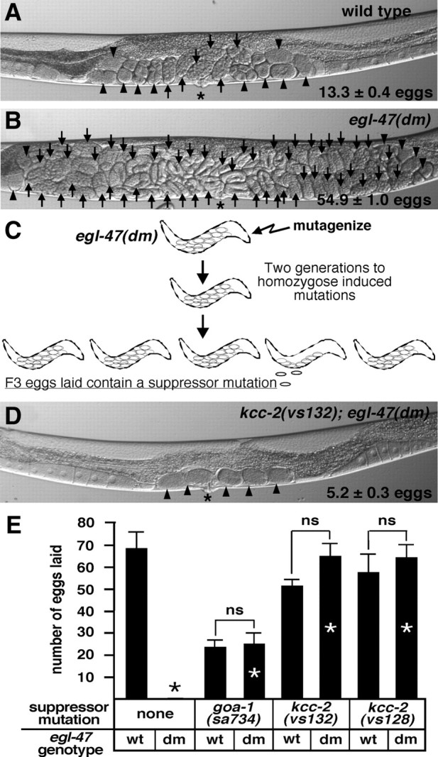
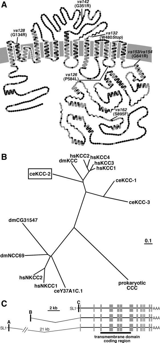
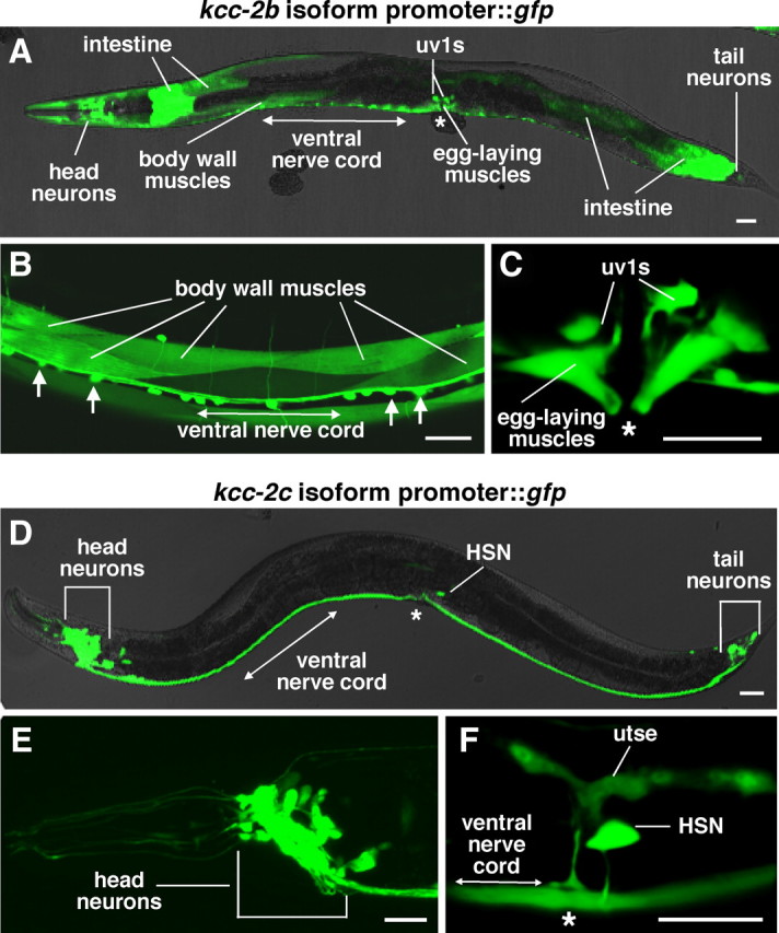
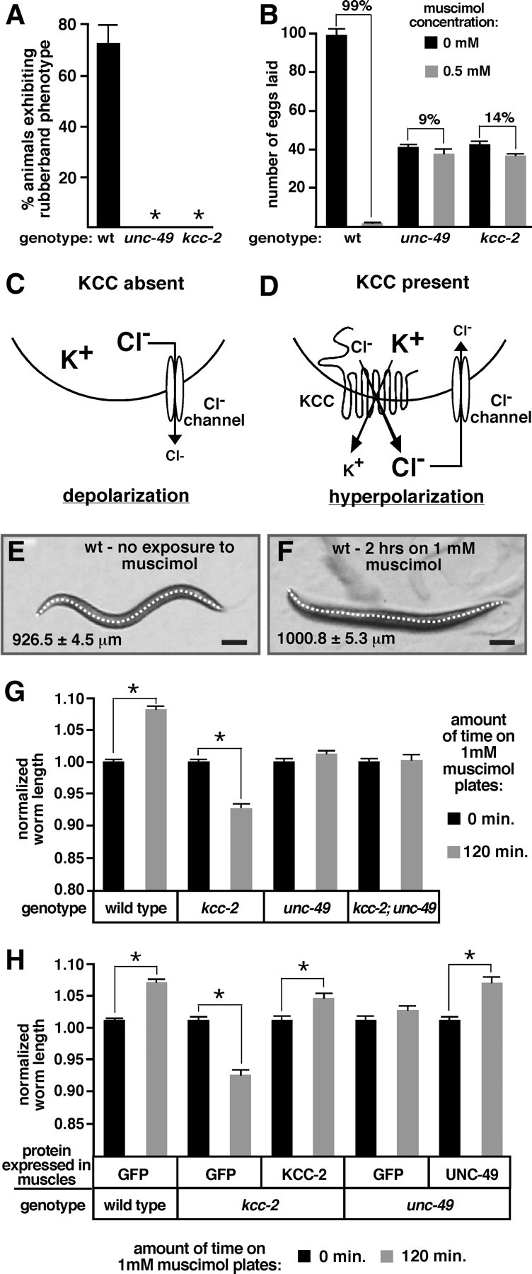
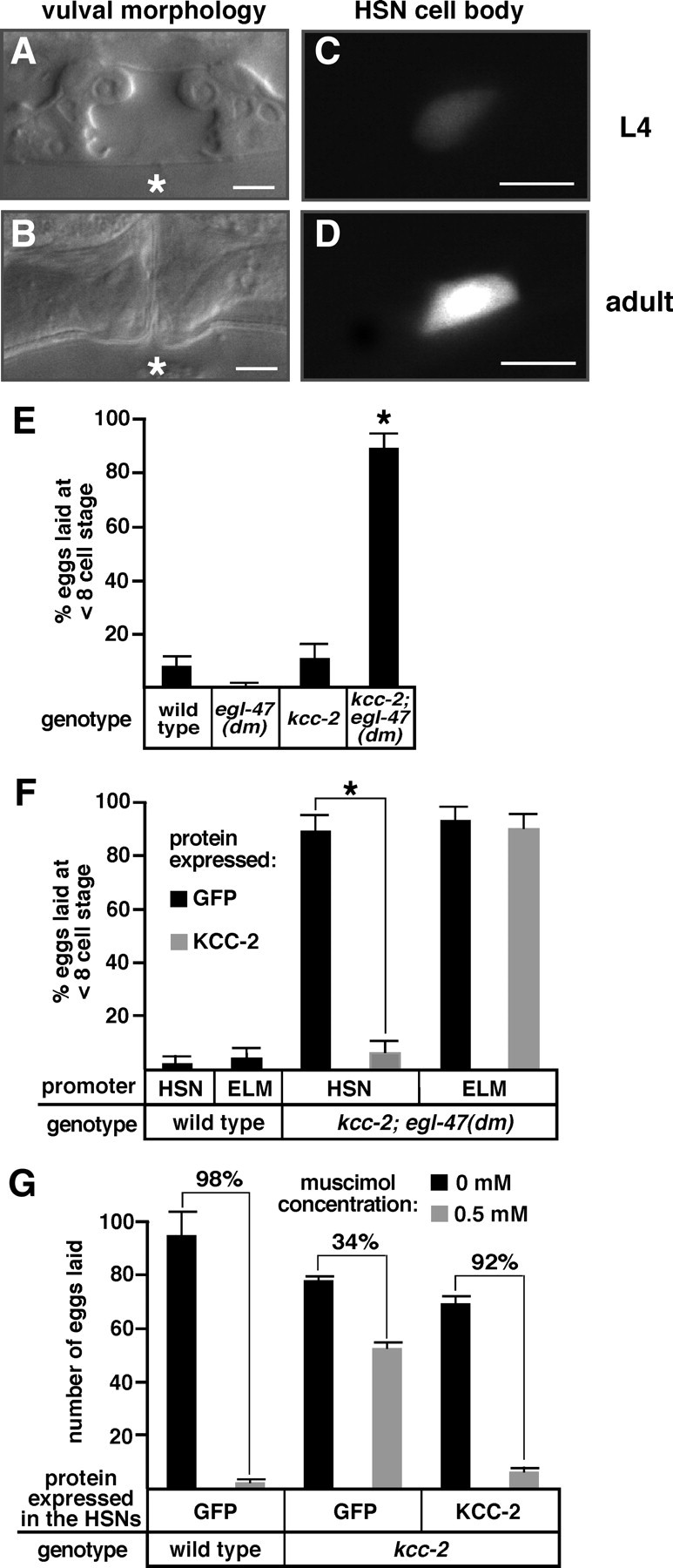
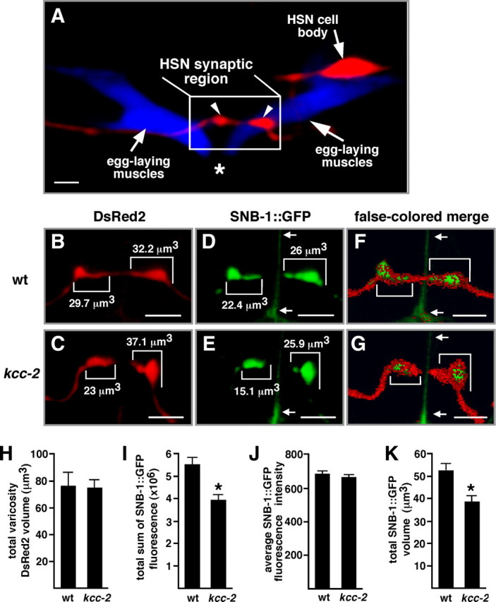
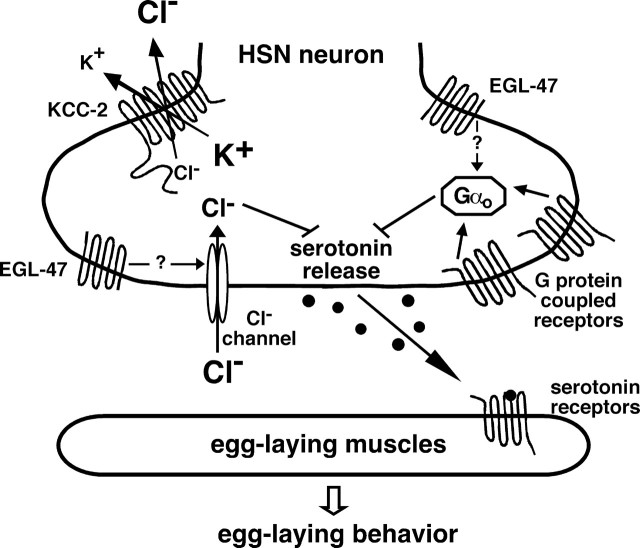
Chloride influx through GABA-gated chloride channels, the primary mechanism by which neural activity is inhibited in the adult mammalian brain, depends on chloride gradients established by the potassium chloride cotransporter KCC2. We used a genetic screen to identify genes important for inhibition of the hermaphrodite-specific motor neurons (HSNs) that stimulate Caenorhabditis elegans egg-laying behavior and discovered mutations in a potassium chloride cotransporter, kcc-2. Functional analysis indicates that, like mammalian KCCs, C. elegans KCC-2 transports chloride, is activated by hypotonic conditions, and is inhibited by the loop diuretic furosemide. KCC-2 appears to establish chloride gradients required for the inhibitory effects of GABA-gated and serotonin-gated chloride channels on C. elegans behavior. In the absence of KCC-2, chloride gradients appear to be altered in neurons and muscles such that normally inhibitory signals become excitatory. kcc-2 is transcriptionally upregulated in the HSN neurons during synapse development. Loss of KCC-2 produces a decrease in the synaptic vesicle population within mature HSN synapses, which apparently compensates for a lack of HSN inhibition, resulting in normal egg-laying behavior. Thus, KCC-2 coordinates the development of inhibitory neurotransmission with synapse maturation to produce mature neural circuits with appropriate activity levels.


Figures

Figure 1.
Figure 1.
Mutations in kcc-2 suppress the egl-47(dm) egg-laying defect. A, B, Representative wild-type and egl-47(dm) animals, respectively. For A, B, and D, the average number of unlaid eggs (±95% confidence interval), eggs with eight cells or fewer (arrowheads), eggs with greater than eight cells (arrows), and the vulva (asterisk) are as indicated. C, A genetic screen was performed to identify suppressors of egl-47(dm). The egl-47(dm) mutant retained many unlaid eggs in utero and never laid any eggs by 18 h after the late L4 stage. egl-47(dm) animals were mutagenized, and F3 eggs laid by the F2 generation 18 h after L4 were picked as suppressors. D, A representative kcc-2(vs132); egl-47(dm) animal. E, By 18 h after L4, the wild type (wt) laid many eggs, whereas the egl-47(dm) mutant never laid an egg. Mutations in goa-1 and kcc-2 suppressed the egl-47(dm) egg-laying defect (compare bars with white asterisks to the black asterisk denoting a measurement of zero eggs). The goa-1 mutant laid fewer eggs than the wild type because the goa-1 mutant produced fewer eggs. The number of eggs laid in E is by five animals by 18 h after L4; error bars show SE. “ns” over brackets indicates measurements that are not significantly different, demonstrating full suppression of egl-47(dm).
Figure 2.
Figure 2.
C. elegans KCC-2 exhibits sequence similarity to mammalian KCCs. A, Predicted transmembrane structure of C. elegans KCC-2 compared with the sequence of rat KCC2. Each amino acid is represented by a circle; gray circles indicate identical amino acids, and black circles represent non-identical amino acids. The locations of the kcc-2 mutations are as indicated. B, Phylogenetic comparison of C. elegans KCC-2 (ceKCC-2; boxed) to the four human KCCs (hsKCC1–hsKCC4), Drosophila melanogaster KCC (dmKCC), two additional C. elegans KCC homologs (ceKCC-1 and ceKCC-3), the human sodium potassium chloride cotransporters (hsNKCC1 and hsNKCC2), two predicted D. melanogaster NKCCs (dmCG31547 and dmNCC69), a predicted C. elegans NKCC (ceY37A1C.1), and a prokaryotic cation chloride cotransporter (CCC). C, Gene structure of C. elegans kcc-2. Three alternative splice forms, kcc-2a, kcc-2b, and kcc-2c, each contain a different 5′ exon (black boxes) that splices onto common exons (gray boxes) encoding the transmembrane domains (black bar). The SL1 trans splice leader (SL1) was identified on the A and C isoforms but not on the B isoform.
Figure 3.
Figure 3.
C. elegans KCC-2 exhibits transport properties similar to those of vertebrate potassium chloride cotransporters. A, B, Xenopus oocytes expressing C. elegans kcc-2, rat Kcc2, or injected with water (none) were tested for uptake of the potassium tracer 86Rb after exposure to hypotonic (Hypo) or isotonic (Iso) solutions containing 79 mm chloride (plus Cl) or 0 mm chloride (no Cl). Measurements in the presence of 250 μm furosemide were used to detect the background from furosemide-insensitive transporters endogenous to Xenopus oocytes. A, C. elegans KCC-2 transport activity was stimulated by hypotonic solution (compare 1 with 7). Isotonic conditions did not stimulate activity (compare 3 with 9). The loop diuretic furosemide inhibited KCC-2 activity (compare 7 with 8). KCC-2 did not uptake 86Rb in the absence of extracellular chloride (compare 7 with 11). Bars 1–6 and 8–12 are significantly different from 7; p < 0.01. Bars 8–12 are not significantly different from the respective water-injected controls (bars 2–6). C. elegans KCC-2 expression resulted in the influx of 2.90 ± 0.51 nmol Rb/h per oocyte. Statistical analysis of non-normalized values resulted in the same significance results. B, Rat KCC2 transport activity was stimulated by hypotonic solution (compare 1 with 7), inhibited by the loop diuretic furosemide (compare 7 with 8), and required extracellular chloride (compare 7 with 11). Rat KCC2 exhibits significant transport under isotonic conditions (compare 3 with 9; p < 0.05), although greater transport activity was observed in hypotonic solution (compare 7 with 9). Bars 1–6 and 8–12 are significantly different from 7; p < 0.01. Expression of rat KCC2 resulted in the influx of 6.96 ± 0.69 nmol Rb/h per oocyte. In A and B, values were normalized to uptake by the transporters under hypotonic conditions plus chloride; error bars show SE. C–F, Oocytes expressing C. elegans KCC-2 (C, E) or rat KCC2 (D, F) were exposed to increasing concentrations of furosemide (C, D) or bumetanide (E, F), from 0 to 250 μm in the 86Rb flux solution. The IC50 value is an average from three experiments, ± SE; C–F show representative experiments. Values were normalized to maximum 86Rb influx in each experiment; error bars indicate SE.
Figure 4.
Figure 4.
C. elegans KCC-2 is predominantly expressed in neurons and muscles. A–C, GFP fluorescence in animals carrying a kcc-2b promoter::gfp::kcc-2 3′ UTR reporter transgene. This reporter transgene was expressed in many cells, including body-wall muscles and ventral cord motor neurons (arrows) (B), as well as egg-laying muscles and uv1 cells (C). D–F, GFP fluorescence in animals carrying a kcc-2c promoter::gfp::kcc-2 3′ UTR reporter transgene. This reporter transgene was expressed in head and tail neurons (E) as well as the HSN neurons and utse (F). Scale bar, 20 μm. Asterisks indicate the position of the vulva.
Figure 5.
Figure 5.
KCC-2 is required for the inhibitory effects of ligand-gated chloride channels. A, Exposure to 1 mm muscimol induced a rubber-band phenotype (quick contraction and relaxation of the body wall muscles after a nose tap) in wild-type (wt) animals but not in unc-49 or kcc-2 mutants. n > 110 for each genotype, error bars show 95% confidence intervals, and asterisks indicate values significantly different from the wild type (p < 0.05). B, The average number of eggs laid by five animals for 2 h on plates in the presence or absence of 0.5 mm muscimol. Animals with mutations in unc-49 and kcc-2 were resistant to the inhibitory effects of muscimol on egg-laying behavior. Brackets are labeled with the percentage inhibition of egg laying induced by muscimol. The unc-49 and kcc-2 mutants retained significantly fewer eggs in utero compared with the wild type as a result of a decrease in egg production and thus laid fewer eggs on the non-muscimol plates. For each genotype and condition, n = 30 animals, and error bars show SE. C, In the absence of KCC activity, intracellular Cl levels remain high, and stimulation of a Cl channel causes Cl to move out of the neuron, leading to depolarization. D, KCCs use the potassium (K+) concentration gradient to drive electroneutral extrusion of K+ and Cl to establish a Cl concentration gradient, with higher extracellular Cl. Activation of a Cl channel allows Cl to reenter the neuron, resulting in neural inhibition. E, F, Representative wild-type animals in the absence (E) and presence (F) of muscimol. A line was drawn down the center of each animal to measure body length. Average body length for each condition is indicated. Scale bars, 100 μm. G, Exposure to muscimol caused a significant increase in body length of wild-type animals, a decrease in the length of the kcc-2 mutant, and no change in the unc-49 single mutant or the kcc-2; unc-49 double mutant. H, After reexpression of kcc-2 in the body-wall muscles of the kcc-2 mutant or reexpression of unc-49 in the body-wall muscles of the unc-49 mutant, muscimol caused a significant increase in body length, as observed in wild-type animals. G, H, For each genotype, measurements were normalized to body length in the absence of muscimol. Error bars show SE, and asterisks indicate a significant difference (p < 0.0001).
Figure 6.
Figure 6.
kcc-2 expression in the HSN neurons is developmentally upregulated and causes a shift in HSN activity. A, B, Bright-field images show L4 and adult vulval morphology of wild-type animals, which was used to precisely determine developmental age. Asterisks indicate the position of the center of the vulva. C, D, GFP expressed by the kcc-2c promoter in HSN cell bodies from the same animals shown in A and B, respectively. Scale bars, 5 μm. E, The kcc-2; egl-47(dm) double mutant laid many early-stage eggs that had not developed beyond the eight-cell stage, whereas such hyperactive egg laying was not seen in the kcc-2 or egl-47(dm) single mutants. F, Reexpression of kcc-2 in the HSN neurons, but not the ELMs of the kcc-2; egl-47(dm) mutant, rescued the synthetic hyperactive egg-laying behavior. For E and F, n ≥ 100 eggs per genotype, error bars show the 95% confidence interval, and asterisks indicate values significantly different from the respective controls (p < 0.05). G, Reexpression of kcc-2 in the HSN neurons of the kcc-2 mutant restored the inhibition of muscimol on egg-laying behavior. Brackets are labeled with the percentage inhibition of egg laying induced by muscimol. For each genotype, n = 50 animals (10 animals from five independent transgenic lines); error bars show SE.
Figure 7.
Figure 7.
kcc-2 mutants exhibit a decrease in the size of the synaptic vesicle population within HSN presynaptic termini. A, Confocal image of the HSNL neuron expressing the red fluorescent protein DsRed2 and the egg-laying muscles expressing the cyan fluorescent protein CFP. The HSN process forms presynaptic varicosities (arrowheads) that contact the egg-laying muscles near the vulva. Scale bar, 5 μm. Asterisk indicates the position of vulva. B, C, Expression of DsRed2 in wild-type (wt) (B) and kcc-2 mutant (C) HSNs. D, E, Expression of SNB-1::GFP in wild-type (D) and kcc-2 mutant (E) HSNs. In B and C, numbers indicate the DsRed2 volume for each varicosity, and, in D and E, numbers indicate the volume of the SNB-1::GFP-labeled structure within each varicosity. Images in B-E are two-dimensional representations of three-dimensional images in which each two-dimensional pixel is shown at the brightest individual voxel intensity from the stack of three-dimensional voxels that it represents. F, G, Merged images in which the SNB-1::GFP is represented as in D and E, but DsRed2 pixels above background that do not overlap with the SNB-1::GFP are false colored red, partially obscuring the green label. These images emphasize that the perimeters of the synaptic varicosities are labeled by DsRed2, and that the SNB-1::GFP-labeled structures are contained within the varicosities. The red-only perimeter is more prominent in kcc-2 than in the wild-type because of the decreased volume of SNB-1::GFP labeling in kcc-2. B–G, Brackets indicate synaptic varicosities, and arrows point to an artifact in the GFP channel caused by the vulval slit. Scale bars, 5 μm. H, No significant difference in total DsRed2 varicosity volume was observed between wild-type and kcc-2 mutant animals (p = 0.89). I, A significant difference in the total sum of SNB-1::GFP fluorescence in the HSN synaptic region was observed between the wild-type and kcc-2 mutant animals (p < 0.005). J, The average SNB-1::GFP fluorescence intensity within the GFP-labeled structures was not significantly different between the wild-type and kcc-2 mutant animals (p = 0.40), suggesting that the density of synaptic vesicles within these structures is unchanged. K, The total volume of SNB-1::GFP-labeled structures in the HSN synaptic region was significantly less in the kcc-2 mutant compared with the wild type (p < 0.005), indicating that the reduction in total SNB-1::GFP fluorescence is likely attributable to a smaller population of synaptic vesicles inside the HSN varicosities of the kcc-2 mutant. For H-K, error bars indicate SE; n = 29 varicosities from 11 animals for the wild type, and n = 36 varicosities from 12 animals for the kcc-2 mutant.
Figure 8.
Figure 8.
Signaling through chloride channels and G-protein-coupled receptors regulates neurotransmitter release from the HSNs. Multiple signals converge on the HSNs and are integrated to set the level of serotonin release to control egg laying. Previous work indicates that the acetylcholine receptor GAR-2 and a serotonin autoreceptor are likely coupled to the Gαo-protein GOA-1 to inhibit neurotransmitter release from the HSNs. Here we demonstrate that KCC-2 acts in the HSNs to establish a chloride gradient necessary for neural inhibition through neurotransmitter-gated chloride channels, which are likely activated by GABA and/or tyramine, to regulate egg laying. Inhibition of HSN activity by EGL-47 depends on both Gαo and KCC-2, allowing genes involved in either process to be identified in the egl-47(dm) suppressor screen. Although EGL-47 is similar to G-protein-coupled receptors in primary structure, it has effects independent of Gαo, and the schematic indicates the possibility that it may activate chloride channels. KCC-2 and Gαo are also expressed in the egg-laying muscles, but both are only shown in the HSNs because this is the only site of function for these proteins in the control of egg laying as identified by cell-specific rescue experiments [this work and the one by Tanis et al. (2008)].

Similar articles

Cited by

References

    1. Adler CE, Fetter RD, Bargmann CI. UNC-6/Netrin induces neuronal asymmetry and defines the site of axon formation. Nat Neurosci. 2006;9:511–518. - PMC - PubMed
    1. Akerman CJ, Cline HT. Depolarizing GABAergic conductances regulate the balance of excitation to inhibition in the developing retinotectal circuit in vivo. J Neurosci. 2006;26:5117–5130. - PMC - PubMed
    1. Bamber BA, Beg AA, Twyman RE, Jorgensen EM. The Caenorhabditis elegans unc-49 locus encodes multiple subunits of a heteromultimeric GABA receptor. J Neurosci. 1999;19:5348–5359. - PMC - PubMed
    1. Bany IA, Dong MQ, Koelle MR. Genetic and cellular basis for acetylcholine inhibition of Caenorhabditis elegans egg-laying behavior. J Neurosci. 2003;23:8060–8069. - PMC - PubMed
    1. Ben-Ari Y. Excitatory actions of gaba during development: the nature of the nurture. Nat Rev Neurosci. 2002;3:728–739. - PubMed

Publication types

MeSH terms

LinkOut - more resources