. 2009 Aug 12;29(32):9967-76.
doi: 10.1523/JNEUROSCI.1151-09.2009.

Overcoming macrophage-mediated axonal dieback following CNS injury

Affiliations

Overcoming macrophage-mediated axonal dieback following CNS injury

Sarah A Busch et al. J Neurosci. .

Abstract

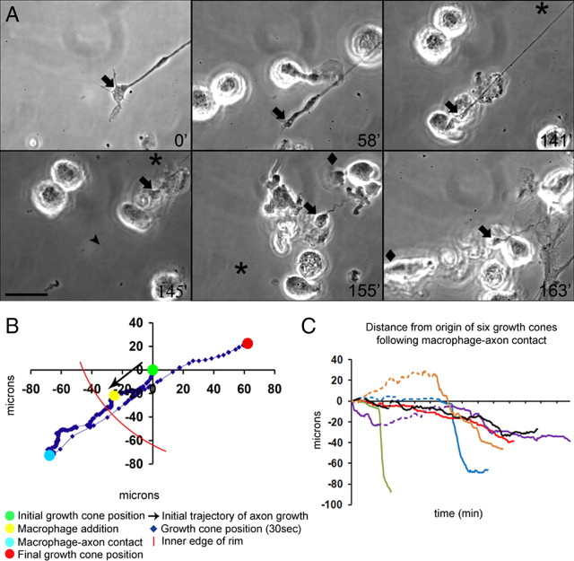
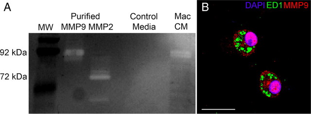
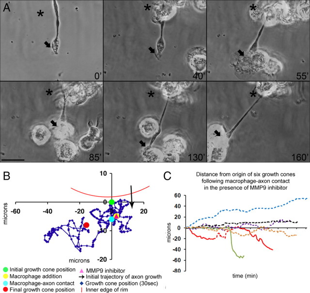
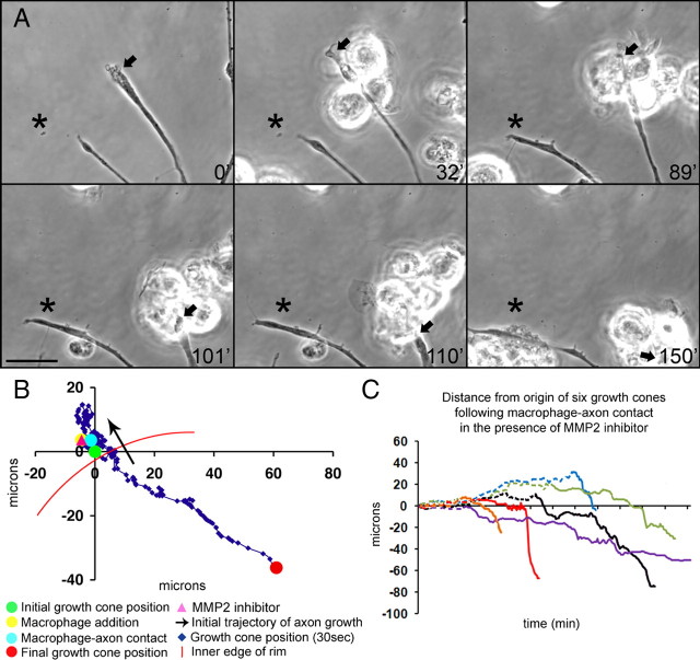
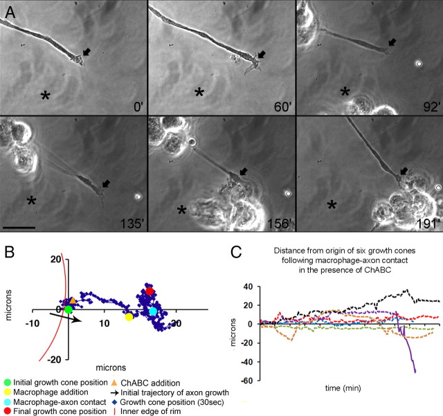
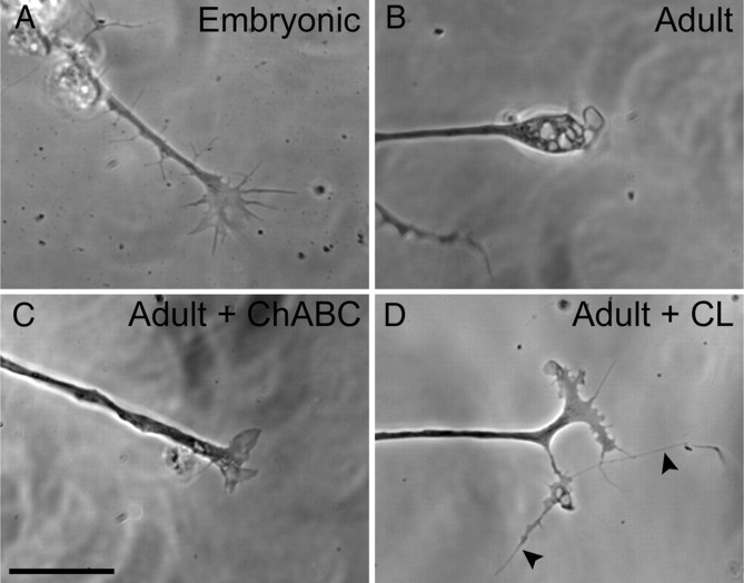
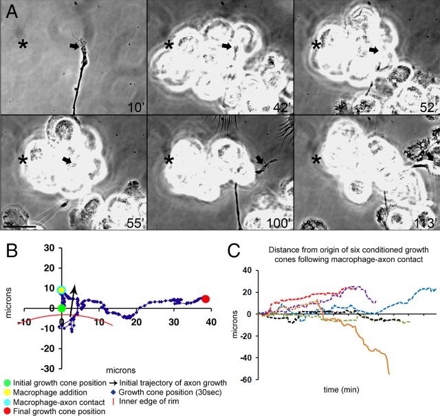
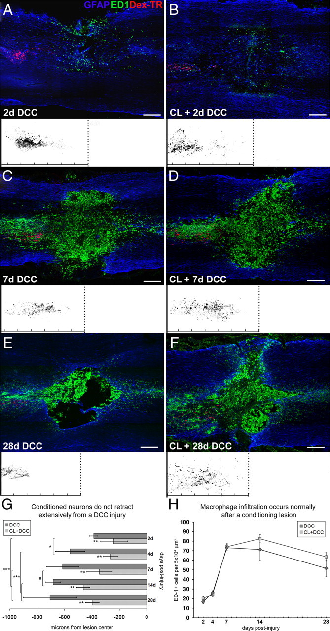
Trauma to the adult CNS initiates multiple processes including primary and secondary axotomy, inflammation, and glial scar formation that have devastating effects on neuronal regeneration. After spinal cord injury, the infiltration of phagocytic macrophages coincides with long-distance axonal retraction from the initial site of injury, a deleterious phenomenon known as axonal dieback. We have previously shown that activated macrophages directly induce long-distance retraction of dystrophic axons in an in vitro model of the glial scar. We hypothesized that treatments that are primarily thought to increase neuronal regeneration following spinal cord injury may in fact derive a portion of their beneficial effects from inhibition of macrophage-mediated axonal retraction. We analyzed the effects of protease inhibition, substrate modification, and neuronal preconditioning on macrophage-axon interactions using our established in vitro model. General inhibition of matrix metalloproteinases and specific inhibition of MMP-9 prevented macrophage-induced axonal retraction despite significant physical interactions between the two cell types, whereas inhibition of MMP-2 had no effect. Chondroitinase ABC-mediated digestion of the aggrecan substrate also prevented macrophage-induced axonal retraction in the presence of extensive macrophage-axon interactions. The use of a conditioning lesion to stimulate intrinsic neuronal growth potential in the absence of substrate modification likewise prevented macrophage-induced axonal retraction in vitro and in vivo following spinal cord injury. These data provide valuable insight into the cellular and molecular mechanisms underlying macrophage-mediated axonal retraction and demonstrate modifications that can alleviate the detrimental effects of this unfavorable phenomenon on the postlesion CNS.


Figures

Figure 1.
Figure 1.
Dystrophic adult dorsal root ganglion axons on an inhibitory proteoglycan gradient retract extensively following macrophage contact. A, Six-panel montage of single frame images extracted from a time-lapse movie. NR8383 macrophages were added to a 2 DIV culture of adult dorsal root ganglion neurons on an inverse spot gradient of growth-promoting molecule laminin and the inhibitory proteoglycan aggrecan. The growth cones imaged were located in the inner edge of the rim, which is approximated as the area into which the majority of satellite cells and neurons cease to extend, leaving a clearing in the region of greatest inhibitory proteoglycan. Times for each frame are given at the bottom right of each image and an asterisk marks a consistent point on the culture dish as a reference point for growth cone position during frame shifts. A diamond indicates a new point of reference in this montage only. An arrow denotes the central domain of the growth cone. Macrophages were added following a 30 min period of observation, and contact occurred at 58 min. In the third frame at 141 min, the growth cone is directly contacted by a macrophage, which immediately precedes the large-scale retraction. Note the presence of a retraction fiber at 145 min (arrowhead). By 163 min, the axon has retracted completely. The entire movie can be viewed as supplemental Movie 1 (available at www.jneurosci.org as supplemental material). B, Positional graph indicating the location of the growth cone for the entire time-lapse movie shown in A. The growth cone retracts a distance of 160 μm at a speed of 8 μm/min. C, Distance from the origin of six dystrophic axons on the aggrecan/laminin spot gradient following contact with macrophages. A dashed line indicates the buffer zone of normal dynamic growth cone extension and collapse, and a solid line indicates a long-distance retraction. Each tick mark on the x-axis represents 5 min. Scale bar: A, 20 μm.
Figure 2.
Figure 2.
Macrophages express and secrete MMP-9 in vitro. A, Gelatin zymographic analysis of macrophage-conditioned media. Zymogram gel indicates the presence of the active form of MMP-9 at 97 kDa in macrophage-conditioned media, but not in nonconditioned control media. There is no evidence of MMP-2 in either macrophage-conditioned or control media. Purified MMP-2 and MMP-9 were used as positive controls. B, Confocal image (100×) of NR8383 macrophages cultured on an inverse spot gradient of proteoglycan and laminin. Immunocytochemistry reveals that activated macrophages, as determined by ED-1 expression (green), contain MMP-9 protein (red). Scale bar: B, 25 μm.
Figure 3.
Figure 3.
Dystrophic adult dorsal root ganglion axons on an inhibitory proteoglycan gradient persist following macrophage contact in the presence of an MMP-9 inhibitor. A, Six-panel montage of a time-lapse movie of NR8383 macrophages and DRG neurons arranged as stated in the Figure 1 legend. Macrophages and MMP-9 inhibitor were added following a 30 min period of observation, and contact occurred at 38 min. Macrophages can be seen physically interacting with the axon at 55 min, 85 min, and 130 min and by 160 min have pulled the growth cone to the far left of the panel, but no retraction is observed. The entire movie can be viewed as supplemental Movie 2 (available at www.jneurosci.org as supplemental material). B, Positional graph indicating the location of the growth cone for the entire time-lapse movie shown in A. C, Distance from the origin of six dystrophic axons on the aggrecan/laminin spot gradient in the presence of an MMP-9 inhibitor following contact with macrophages. A dashed line indicates the buffer zone of normal dynamic growth cone extension and collapse and a solid line indicates a long-distance retraction. Each tick mark on the x-axis represents 5 min. Scale bar: A, 20 μm.
Figure 4.
Figure 4.
Dystrophic adult dorsal root ganglion axons on an inhibitory proteoglycan gradient retract following macrophage contact in the presence of an MMP-2 inhibitor. A, Six-panel montage of a time-lapse movie of NR8383 macrophages and DRG neurons arranged as stated in the Figure 1 legend. Macrophages and MMP-2 inhibitor were added following a 30 min period of observation, and contact occurred at 32 min. Axonal retraction can be seen at 101 min and has completed by 150 min. The entire movie can be viewed as supplemental Movie 3 (available at www.jneurosci.org as supplemental material). B, Positional graph indicating the location of the growth cone for the entire time-lapse movie shown in A. C, Distance from the origin of six dystrophic axons on the aggrecan/laminin spot gradient in the presence of an MMP-2 inhibitor following contact with macrophages. A dashed line indicates the buffer zone of normal dynamic growth cone extension and collapse, and a solid line indicates a long-distance retraction. Each tick mark on the x-axis represents 5 min. Scale bar: A, 20 μm.
Figure 5.
Figure 5.
Dystrophic adult dorsal root ganglion axons on an inhibitory proteoglycan gradient treated with ChABC persist after macrophage contact. A, Six-panel montage of a time-lapse movie of NR8383 macrophages and DRG neurons arranged as stated in the Figure 1 legend. ChABC (0.1 U/ml) was added to the dish following a 30 min period of observation and growth cones were observed for positional and morphological changes for 30 min before macrophage addition. Macrophage contact occurred at 62 min, and the axon did not retract by the end of time-lapse imaging at 180 min. B, Positional graph indicating the location of the growth cone for the entire time-lapse movie shown in A. Note the forward movement of the growth cone into the rim after ChABC addition. C, Distance from the origin of six dystrophic axons on the aggrecan/laminin spot gradient treated with ChABC following contact with macrophages. A dashed line indicates the buffer zone of normal dynamic growth cone extension and collapse and a solid line indicates a long-distance retraction. Each tick mark on the x-axis represents 5 min. Scale bar: A, 20 μm.
Figure 6.
Figure 6.
Comparison of embryonic, dystrophic, ChABC treated, and conditioned adult growth cones. A, Growth cone of an embryonic day 16 dorsal root ganglion neuron on an inverse spot gradient of growth-promoting molecule laminin and the inhibitory proteoglycan aggrecan. The growth cone possesses numerous filopodia extending from a broad central lamellipodium. B, Growth cone of a dystrophic adult dorsal root ganglion neuron on an inverse spot gradient of growth-promoting molecule laminin and the inhibitory proteoglycan aggrecan. This dystrophic growth cone appears club-like without filopodia and lamellipodia and contains numerous vesicles. C, Growth cone of an adult dorsal root ganglion neuron following treatment with ChABC on an inverse spot gradient of growth-promoting molecule laminin and the inhibitory proteoglycan aggrecan. The growth cone has responded to ChABC treatment of the substrate by elaborating a wide lamellipodium. D, Growth cone of a conditioned adult dorsal root ganglion neuron on an inverse spot gradient of growth-promoting molecule laminin and the inhibitory proteoglycan aggrecan. The conditioned growth cone has lengthy filopodia extending from a broad lamellipodium. Arrows indicate extremely long filopodia. It is important to note that both substrate modification (C) and conditioning lesion (D) induce growth cone structures that are reminiscent of embryonic neurons. Scale bar: 20 μm.
Figure 7.
Figure 7.
Conditioned adult dorsal root ganglion axons on an inhibitory proteoglycan gradient persist after macrophage contact. A, Six-panel montage of a time-lapse movie of NR8383 macrophages and conditioned DRG neurons arranged as stated in the Figure 1 legend. Macrophage contact occurred at 42 min and the axon did not retract by the end of time-lapse imaging. The entire movie can be viewed as supplemental Movie 4 (available at www.jneurosci.org as supplemental material). B, Positional graph indicating the location of the growth cone for the entire time-lapse movie shown in A. Note the lateral movement of the growth cone along the rim. C, Distance from the origin of six axons of conditioned neurons on the aggrecan/laminin spot gradient following contact with macrophages. A dashed line indicates the buffer zone of normal dynamic growth cone extension and collapse, and a solid line indicates a long-distance retraction. Each tick mark on the x-axis represents 5 min. Scale bar: A, 20 μm.
Figure 8.
Figure 8.
Conditioned neurons persist close to the lesion center after spinal cord injury. A–F, Confocal montages (10×) of longitudinal sections of animals receiving a dorsal column crush (DCC) spinal cord injury alone or a dorsal column crush spinal cord injury 7 d after receiving a sciatic crush conditioning lesion (CL + DCC). Caudal is on the left side of the image and rostral is on the right. Dorsal root ganglion neuron are labeled with Texas Red-conjugated dextran 3000 MW (Dex-TR) (red) and ED-1+ macrophages/microglia (green) and GFAP+ astrocytes (blue). Below each confocal image are superimposed fiber tracings of three sections from a representative animal for each time point. Dotted lines represent the lesion center, and ruler tick marks are 200 μm each. A, At 2 d after DCC, sensory axons of the dorsal columns have retracted a short distance from the center of the lesion, determined by GFAP+ astrocytes (blue). Few ED-1+ macrophages/microglia (green) are present in the lesion at this time. B, At 2 d after CL + DCC, sensory axons have retracted a short distance from the lesion center, similar to the level observed in DCC animals. C, At 7 d after DCC, numerous ED-1+ phagocytic cells are present within the lesion and sensory axons have retracted a significant distance from the lesion center. D, At 7 d after CL + DCC, fibers are still in close proximity to the center of the lesion despite the presence of numerous ED-1+ phagocytic cells. E, At 28 d after DCC, axons have retracted a considerable distance from the lesion center. F, At 28 d after DCC plus conditioning lesion 1 week before DCC lesion, axons persist close to the lesion center despite the presence of numerous ED-1+ cells, and the fiber front is wider than in control DCC animals. G, Quantification of axonal retraction from the center of the lesion over time. All days are significantly different from one another by one-way ANOVA, F(4,440) = 28.71. Day 2 is significantly different from days 4, 7, 14, and 28; day 4 is significantly different from days 14 and 28; day 7 is significantly different from day 14 by Tukey's post hoc test, #p < 0.01, *p < 0.005, ***p < 0.00005. Control and conditioning lesion groups are significantly different from one another (one-way ANOVA, F(1,440) = 195.47, Tukey's post hoc test, **p < 0.0005). Error bars indicate SEM. H, Macrophage infiltration is not significantly different in control and conditioned animals. Scale bars: A–F, 250 μm.

Similar articles

Cited by

References

    1. Baba T, Iwasaki S, Maruoka T, Suzuki A, Tomaru U, Ikeda H, Yoshiki T, Kasahara M, Ishizu A. Rat CD4+CD8+ macrophages kill tumor cells through an NKG2D- and granzyme/perforin-dependent mechanism. J Immunol. 2008;180:2999–3006. - PubMed
    1. Barritt AW, Davies M, Marchand F, Hartley R, Grist J, Yip P, McMahon SB, Bradbury EJ. Chondroitinase ABC promotes sprouting of intact and injured spinal systems after spinal cord injury. J Neurosci. 2006;26:10856–10867. - PMC - PubMed
    1. Bradbury EJ, Moon LD, Popat RJ, King VR, Bennett GS, Patel PN, Fawcett JW, McMahon SB. Chondroitinase ABC promotes functional recovery after spinal cord injury. Nature. 2002;416:636–640. - PubMed
    1. Busch SA, Silver J. The role of extracellular matrix in CNS regeneration. Curr Opin Neurobiol. 2007;17:120–127. - PubMed
    1. Chandler S, Miller KM, Clements JM, Lury J, Corkill D, Anthony DC, Adams SE, Gearing AJ. Matrix metalloproteinases, tumor necrosis factor and multiple sclerosis: an overview. J Neuroimmunol. 1997;72:155–161. - PubMed

Publication types

MeSH terms