. 2009 Aug 12;29(32):10104-10.
doi: 10.1523/JNEUROSCI.2087-09.2009.

Glycinergic projection neurons of the cerebellum

Affiliations

Glycinergic projection neurons of the cerebellum

Martha W Bagnall et al. J Neurosci. .

Abstract

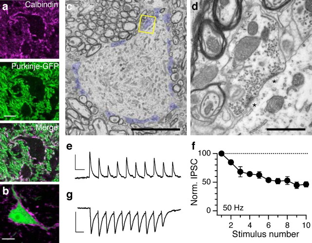
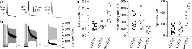
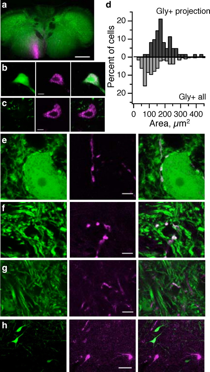
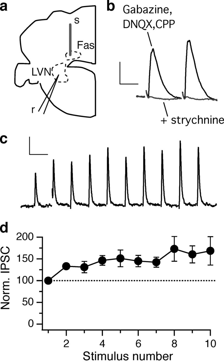
The cerebellum funnels its entire output through a small number of presumed glutamatergic premotor projection neurons in the deep cerebellar nuclei and GABAergic neurons that feed back to the inferior olive. Here we use transgenic mice selectively expressing green fluorescent protein in glycinergic neurons to demonstrate that many premotor output neurons in the medial cerebellar (fastigial) nuclei are in fact glycinergic, not glutamatergic as previously thought. These neurons exhibit similar firing properties as neighboring glutamatergic neurons and receive direct input from both Purkinje cells and excitatory fibers. Glycinergic fastigial neurons make functional projections to vestibular and reticular neurons in the ipsilateral brainstem, whereas their glutamatergic counterparts project contralaterally. Together, these data suggest that the cerebellum can influence motor outputs via two distinct and complementary pathways.


Figures

Figure 1.
Figure 1.
Identification of large glycinergic neurons in the fastigial nuclei. a, Coronal section of cerebellum and brainstem in the GlyT2–GFP mouse. Large GFP+ cells are evident in the fastigial (Fas) but not the interpositus (Int) or dentate (Den) deep cerebellar nuclei. Scattered cerebellar Golgi cells are also visible. IV, Fourth ventricle. Scale bar, 500 μm. b, Confocal image of a large fastigial GFP+ neuron; c, small dentate GFP+ presumed interneurons. Images represent several averaged z-planes totaling <3 μm. Scale bars, 10 μm. d, Single-cell RT-PCR reveals that large fastigial GFP neurons express the glutamatergic marker VGluT2, whereas GFP+ neurons express GlyT2. Right, Whole-brain RNA processed alongside experimental samples. Bottom ladder band is 200 bp, with increments of 100 bp. Performed as in the study by Bagnall et al. (2007).
Figure 2.
Figure 2.
Purkinje cells inhibit large fastigial glycinergic neurons. a, Calbindin immunostaining (top) identifies Purkinje cell terminals, visualized in the L7–GFP mouse (center) (Sekirnjak et al., 2003). Scale bar, 10 μm. b, Calbindin immunostaining (magenta) in the GlyT2–GFP line reveals that large fastigial glycinergic neurons are surrounded by Purkinje cell terminals. All confocal images represent one to six averaged z-sections (<3 μm total). Scale bar, 10 μm. c, Electron micrograph of a large glycinergic neuron showing many Purkinje cell synapses (blue) on soma. Scale bar, 10 μm. d, Higher-magnification view of the synapse outlined in c with two release sites (asterisks) displaying typical Purkinje cell features: large bouton size, symmetric synaptic density, and elliptical vesicles. Scale bar, 1 μm. e, White matter stimulation during whole-cell recording of large glycinergic fastigial neuron elicits a gabazine-sensitive synaptic current. Stimulus artifacts blanked for clarity. Calibration: 100 pA, 20 ms. f, GABAergic currents exhibit sustained transmission at 50 Hz (n = 4; mean ± SEM). g, Glutamatergic synaptic currents (DNQX- and CPP-sensitive) recorded in another large glycinergic fastigial neuron. Calibration: 100 pA, 20 ms.
Figure 3.
Figure 3.
Physiological characteristics of large fastigial glycinergic neurons resemble those of large glutamatergic neurons, not small glycinergic neurons. a, Action potential waveforms recorded from a typical large glycinergic neuron (left), large glutamatergic neuron (middle), and small glycinergic neuron (right). b, Response to a 1 s step of depolarizing current at the maximum level to which the neuron could fire continuously across the whole step. Right axis, Instantaneous firing rate. c, Small glycinergic neurons are significantly different from both large glutamatergic and large glycinergic neurons in action potential width (left), maximum firing rate (middle), and input resistance (right) (p < 0.05, Wilcoxon's unpaired t test, for all small vs large comparisons), whereas large glutamatergic and glycinergic neurons did not differ significantly (p > 0.3, all large glycinergic vs large glutamatergic comparisons).
Figure 4.
Figure 4.
Glycinergic fastigial neurons project to the ipsilateral brainstem, whereas glutamatergic neurons project contralaterally. a, Injection site in caudal medulla. Scale bar, 500 μm. b, Ipsilateral to injection, retrogradely labeled fastigial neurons are glycinergic (GFP+). In this and subsequent images: left, GFP expression; middle, retrograde labeling; right, merge. Scale bar, 10 μm. c, Contralateral to injection, retrogradely labeled fastigial neurons are glutamatergic. d, Histogram distribution of somatic area of retrogradely labeled Gly+ cells (top; n = 61 neurons from several animals) versus all Gly+ deep nuclear neurons (bottom; n = 132 neurons). e, f, Anterogradely labeled glycinergic fastigial axons synapse onto glycinergic (e) and non-glycinergic (f) neurons in the ventromedial medullary reticular formation. g, Axonal swellings from glycinergic fastigial projections are also seen in the vestibular nuclei (here, the spinal vestibular nucleus). h, No glycinergic retrogradely labeled neurons were seen after tracer injections to the thalamus. Scale bar, 50 μm.
Figure 5.
Figure 5.
Functional inhibitory projection from the fastigial nucleus to the brainstem. a, Diagram of the recording setup. Left half of a coronal section; top is dorsal, right is medial. A concentric bipolar stimulating electrode (s) was placed in the fastigial nucleus (Fas), and whole-cell recordings (r) were made in the lateral vestibular nucleus (LVN). b, The outward synaptic current recorded in a lateral vestibular nucleus neuron in the presence of ionotropic GABAergic and glutamatergic antagonists is abolished by application of strychnine (1 μm). Calibration: 100 pA, 10 ms. Stimulus artifacts have been blanked. c, Response to a stimulus train at 50 Hz in another lateral vestibular nucleus neuron. The subtracted strychnine-sensitive component is shown. Calibration: 100 pA, 20 ms. d, Fastigial glycinergic currents facilitated during a train of 10 stimuli delivered at 50 Hz (n = 5; mean ± SEM).

Similar articles

Cited by

References

    1. Aizenman CD, Huang EJ, Manis PB, Linden DJ. Use-dependent changes in synaptic strength at the Purkinje cell to deep nuclear synapse. Prog Brain Res. 2000;124:257–273. - PubMed
    1. Aizenman CD, Huang EJ, Linden DJ. Morphological correlates of intrinsic electrical excitability in neurons of the deep cerebellar nuclei. J Neurophysiol. 2003;89:1738–1747. - PubMed
    1. Angaut P, Bowsher D. Ascending projections of the medial cerebellar (fastigial) nucleus: an experimental study in the cat. Brain Res. 1970;24:49–68. - PubMed
    1. Asanuma C, Thach WT, Jones EG. Brainstem and spinal projections of the deep cerebellar nuclei in the monkey, with observations on the brainstem projections of the dorsal column nuclei. Brain Res. 1983;286:299–322. - PubMed
    1. Bagnall MW, Stevens RJ, du Lac S. Transgenic mouse lines subdivide medial vestibular nucleus neurons into discrete, neurochemically distinct populations. J Neurosci. 2007;27:2318–2330. - PMC - PubMed

Publication types

MeSH terms

LinkOut - more resources