. 2009 Aug 12;29(32):10144-52.
doi: 10.1523/JNEUROSCI.1856-09.2009.

Age-dependent impairment of spine morphology and synaptic plasticity in hippocampal CA1 neurons of a presenilin 1 transgenic mouse model of Alzheimer's disease

Affiliations

Age-dependent impairment of spine morphology and synaptic plasticity in hippocampal CA1 neurons of a presenilin 1 transgenic mouse model of Alzheimer's disease

Alexandra Auffret et al. J Neurosci. .

Abstract

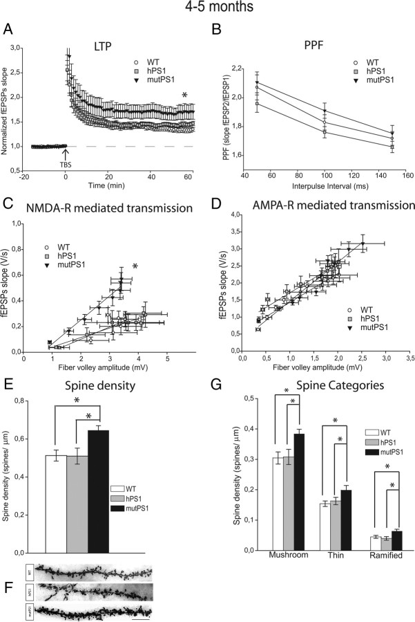
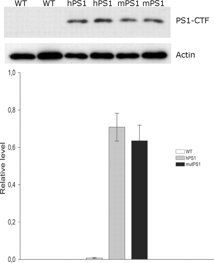
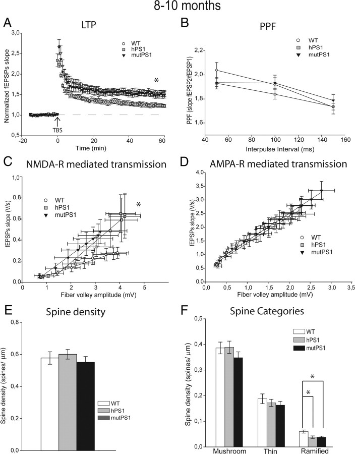
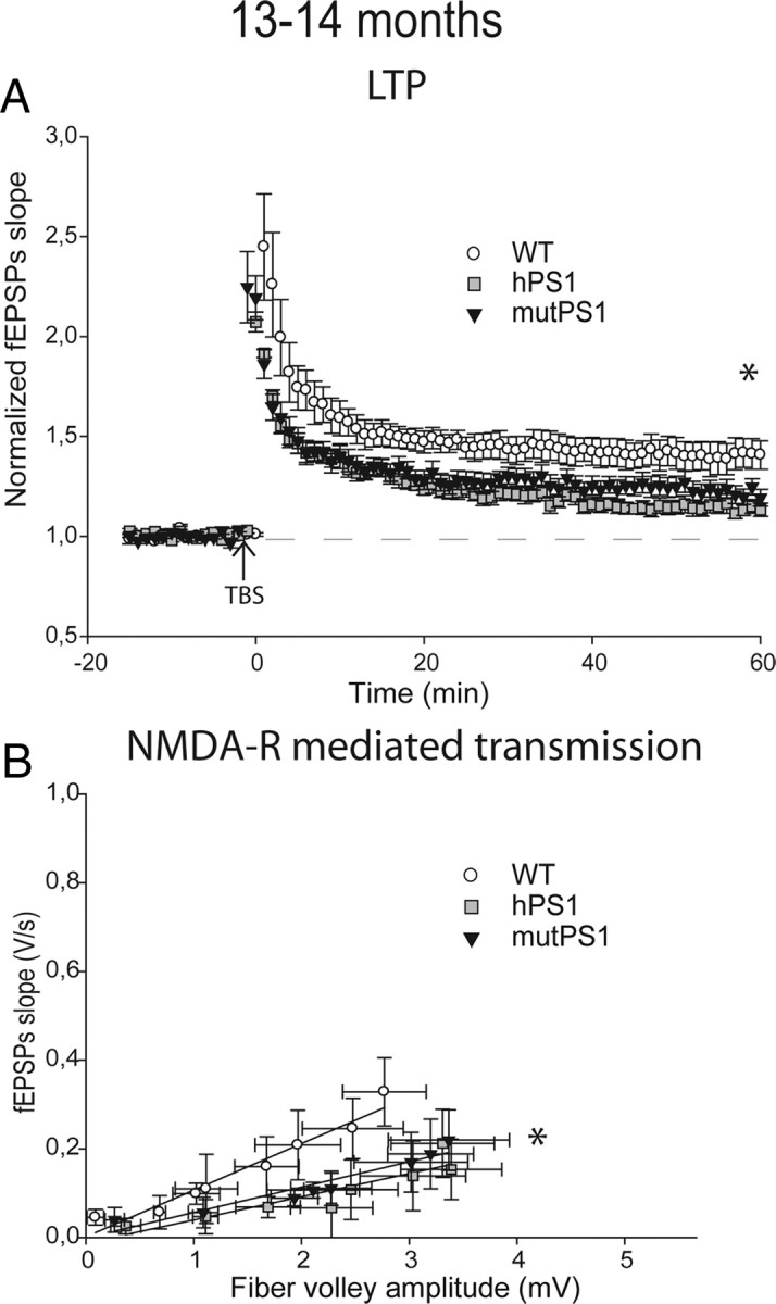
Presenilin 1 (PS1) mutations are responsible for a majority of early onset familial Alzheimer's disease (FAD) cases, in part by increasing the production of Abeta peptides. However, emerging evidence suggests other possible effects of PS1 on synaptic dysfunction where PS1 might contribute to the pathology independent of Abeta. We chose to study the L286V mutation, an aggressive FAD mutation which has never been analyzed at the electrophysiological and morphological levels. In addition, we analyzed for the first time the long term effects of wild-type human PS1 overexpression. We investigated the consequences of the overexpression of either wild-type human PS1 (hPS1) or the L286V mutated PS1 variant (mutPS1) on synaptic functions by analyzing synaptic plasticity and associated spine density changes from 3 to 15 months of age. We found that mutPS1 induces a transient increase observed only in 4- to 5-month-old mutPS1 animals in NMDA receptor (NMDA-R)-mediated responses and LTP compared with hPS1 mice and nontransgenic littermates. The increase in synaptic functions is concomitant with an increase in spine density. With increasing age, however, we found that the overexpression of human wild-type PS1 progressively decreased NMDA-R-mediated synaptic transmission and LTP, without neurodegeneration. These results identify for the first time a transient increase in synaptic function associated with L286V mutated PS1 variant in an age-dependent manner. In addition, they support the view that the PS1 overexpression promotes synaptic dysfunction in an Abeta-independent manner and underline the crucial role of PS1 during both normal and pathological aging.


Figures

Figure 1.
Figure 1.
NMDA-R-mediated transmission, LTP, and spine density are increased by mutPS1 at 4–5 months of age. A, At 4–5 months, the LTP protocol induced a significantly larger response in mutPS1 mice than in WT mice and hPS1 mice (p < 0.05). No significant difference was observed between WT and hPS1 mice. B, PPF was significantly unchanged at 4–5 months of age. C, At 4–5 months, analysis of input-output slopes demonstrates a significant increase of NMDA receptor-mediated responses in mutPS1 compared with hPS1 and WT littermates (p < 0.05). D, No significant difference was found between the three groups of mice at any stimulus level examined for averaged input-output plots of AMPA receptor-mediated responses. E, Quantification of spine density revealed a significant increase (27%) in mutPS1 at 4–5 months of age relative to hPS1 and WT mice (*p < 0.05; Tukey's test). F, Representative segments from 4 to 5-month-old WT, hPS1, and mutPS1 mice. Segments are from the stratum radiatum layer. Scale bar, 10 μm. A Z-stack of photomicrographs was obtained with a Zeiss Axioplan microscope using 100× magnification and oil immersion, and projected on a plane using the MIP method. G, At 4–5 months of age, a significant increase in overall spine density in mutPS1 mice was the result of increases in all three categories of spines (*p < 0.05; Tukey's test).
Figure 2.
Figure 2.
Similar PS1 overexpression levels in hPS1 and mutPS1 mice. Overexpression of human PS1 transgenes was examined in WT, hPS1, and mutPS1 mice (10 months of age) using Western blot of membrane protein extracts from the cerebral cortex. The human PS1 transgene is absent in WT mice and present at the same level in hPS1 and mutPS1 mice, as detected with using anti-C-terminal fragment of human PS1. Results were quantified and PS1 levels were normalized to β-actin levels (n = 3 animals per genotype), and statistical analysis (Mann–Whitney test) shows no difference between hPS1 and mutPS1 mice.
Figure 3.
Figure 3.
Overexpression effects of PS1 during aging. A, At 8–10 months of age, LTP was markedly impaired in hPS1 mice (p < 0.05). In contrast, LTP in the WT mice was normal and similar to that observed in the mutPS1 mice. B, PPF was unchanged in different genotypes analyzed. C, At 8–10 months, analysis of input-output slopes demonstrates a significant increase of basal synaptic transmission in mutPS1 and hPS1 compared with WT mice, indicating enhanced NMDA receptor-mediated synaptic transmission (p < 0.05, Kruskal–Wallis test). D, At 8–10 months of age, no statistically significant difference was detected for averaged input-output plots of AMPA receptor-mediated responses. E, The spine density increase observed at 4–5 months of age in mutPS1 mice did not persist at 8–10 months of age. No statistically significant difference in total spine density at 8–10 months of age was detected between WT, hPS1, and mutPS1 mice. F, At 8–10 months of age, hPS1 and mutPS1 mice showed a significant reduction in ramified spine density (33%) compared with WT mice (*p < 0.05; Tukey's test). If we compare ages inside each genotype for each type of spines, in mutPS1 only, the ramified spine density decreases significantly (33%) at 8–10 months of age compared with 4–5 months of age. This was confirmed by a significant interaction between age and genotype in a two way ANOVA.
Figure 4.
Figure 4.
NMDA-R-mediated transmission and LTP are decreased at 13–14 months of age. A, LTP is markedly impaired in hPS1 mice and mutPS1 compared with WT animals (p < 0.05). In contrast, LTP in the WT mice was normal and similar to that observed at young ages. B, At 13–14 months, analysis of input-output slopes demonstrates a significant decrease of basal synaptic transmission in mutPS1 and hPS1 compared with WT mice, indicating an alteration of NMDA receptor-mediated synaptic transmission compared with young ages (p < 0.05, Kruskal–Wallis test).
Figure 5.
Figure 5.
Synaptic dysfunction are not associated with neuronal death. Dark staining indicates DNA fragmentation induced by DNase treatment of hippocampus (A) and dentate gyrus (B). Dark staining indicates apoptotic germ cells of mouse testis, a second positive control for TUNEL staining (C). Signal is absent in WT, hPS1, and mutPS1 mice in hippocampus (D–F) and particularly in the CA1 region (D1, E1, F1) at 15 months of age. Scale bars, 200 and 20 μm.

Similar articles

Cited by

References

    1. Alvarez VA, Sabatini BL. Anatomical and physiological plasticity of dendritic spines. Annu Rev Neurosci. 2007;30:79–97. - PubMed
    1. Barrow PA, Empson RM, Gladwell SJ, Anderson CM, Killick R, Yu X, Jefferys JG, Duff K. Functional phenotype in transgenic mice expressing mutant human presenilin-1. Neurobiol Dis. 2000;7:119–126. - PubMed
    1. Bonny C, Oberson A, Negri S, Sauser C, Schorderet DF. Cell-permeable peptide inhibitors of JNK: novel blockers of beta-cell death. Diabetes. 2001;50:77–82. - PubMed
    1. Chang EH, Savage MJ, Flood DG, Thomas JM, Levy RB, Mahadomrongkul V, Shirao T, Aoki C, Huerta PT. AMPA receptor downscaling at the onset of Alzheimer's disease pathology in double knockin mice. Proc Natl Acad Sci U S A. 2006;103:3410–3415. - PMC - PubMed
    1. Citron M, Westaway D, Xia W, Carlson G, Diehl T, Levesque G, Johnson-Wood K, Lee M, Seubert P, Davis A, Kholodenko D, Motter R, Sherrington R, Perry B, Yao H, Strome R, Lieberburg I, Rommens J, Kim S, Schenk D, et al. Mutant presenilins of Alzheimer's disease increase production of 42-residue amyloid beta-protein in both transfected cells and transgenic mice. Nat Med. 1997;3:67–72. - PubMed

Publication types

MeSH terms

Substances