. 2009 Oct;102(4):2208-19.
doi: 10.1152/jn.00520.2009. Epub 2009 Aug 12.

Activity-dependent modulation of glutamatergic signaling in the developing rat dorsal horn by early tissue injury

Affiliations

Activity-dependent modulation of glutamatergic signaling in the developing rat dorsal horn by early tissue injury

Jie Li et al. J Neurophysiol. 2009 Oct.

Abstract

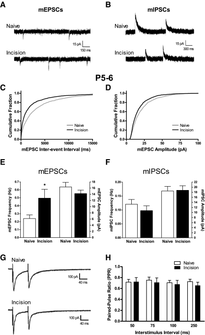
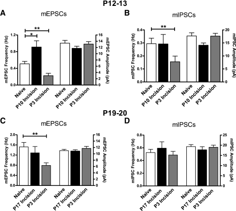
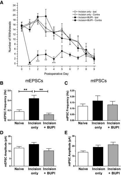
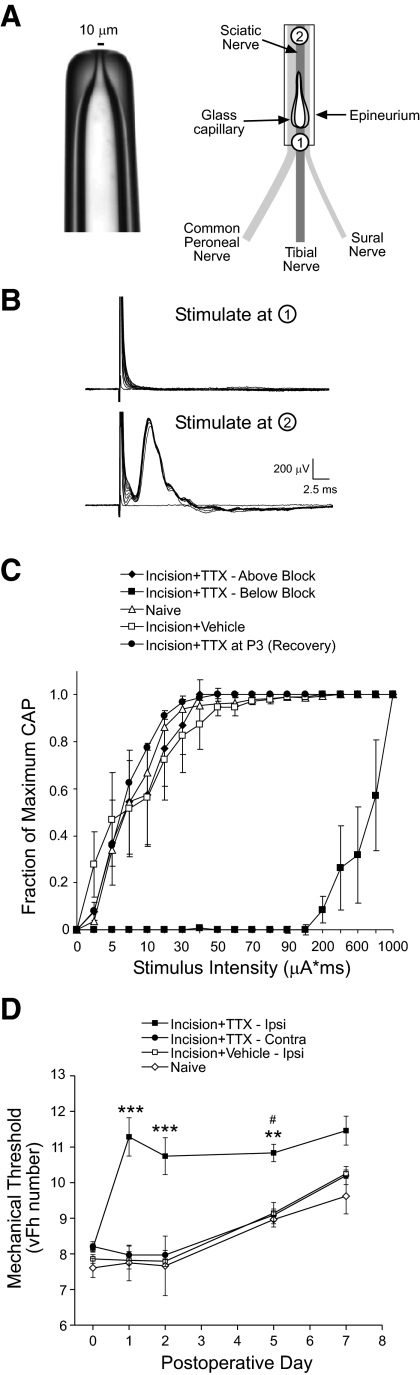
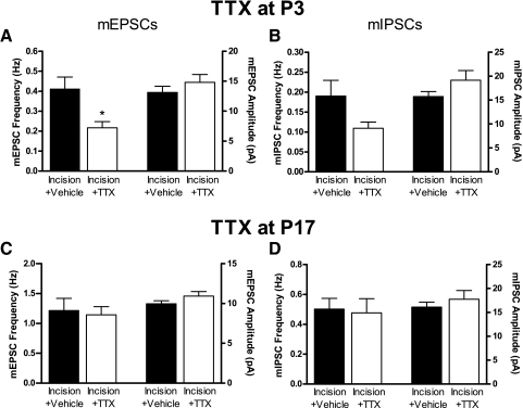
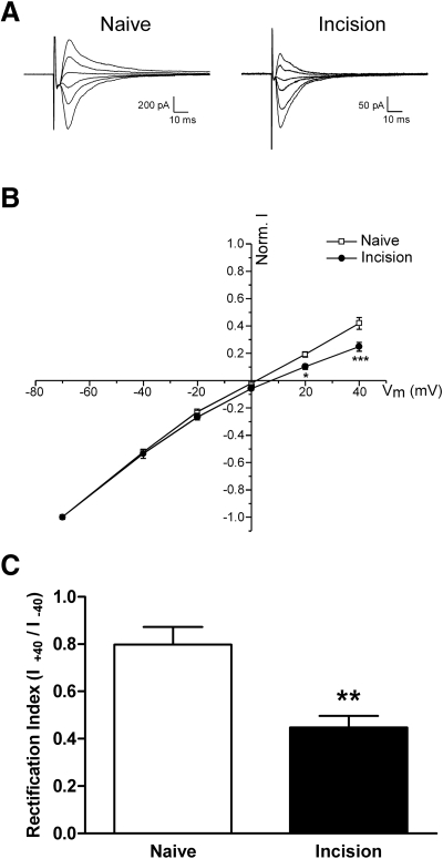
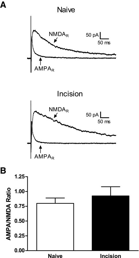
Tissue injury in early life can produce distinctive effects on pain processing, but little is known about the underlying neural mechanisms. Neonatal inflammation modulates excitatory synapses in spinal nociceptive circuits, but it is unclear whether this results directly from altered afferent input. Here we investigate excitatory and inhibitory synaptic transmission in the rat superficial dorsal horn following neonatal hindlimb surgical incision using in vitro patch-clamp recordings and test the effect of blocking peripheral nerve activity on the injury-evoked changes. Surgical incision through the skin and muscle of the hindlimb at postnatal day 3 (P3) or P10 selectively increased the frequency, but not amplitude, of glutamatergic miniature excitatory postsynaptic currents (mEPSCs) recorded 2-3 days after injury, without altering miniature inhibitory postsynaptic current frequency or amplitude at this time point. Meanwhile, incision at P17 failed to affect excitatory or inhibitory synaptic function at 2-3 days postinjury. The elevated mEPSC frequency was accompanied by increased inward rectification of evoked alpha-amino-3-hydroxy-5-methyl-4-isoxazolepropionic acid receptor (AMPAR)-mediated currents, but no change in AMPAR/N-methyl-D-aspartate receptor ratios, and was followed by a persistent reduction in mEPSC frequency by 9-10 days postinjury. Prolonged blockade of primary afferent input from the time of injury was achieved by administration of bupivacaine hydroxide or tetrodotoxin to the sciatic nerve at P3. The increase in mEPSC frequency evoked by P3 incision was prevented by blocking sciatic nerve activity. These results demonstrate that increased afferent input associated with peripheral tissue injury selectively modulates excitatory synaptic drive onto developing spinal sensory neurons and that the enhanced glutamatergic signaling in the dorsal horn following neonatal surgical incision is activity dependent.


Figures

Fig. 1.
Fig. 1.
Surgical incision during early life potentiates excitatory synaptic transmission in the developing rat superficial dorsal horn (SDH) at 2–3 days postinjury. A: examples of miniature excitatory postsynaptic currents (mEPSCs) recorded at a holding potential (Vh) of −70 mV in postnatal day 5 (P5) to P6 SDH neurons from naïve pups or littermates undergoing surgical incision at P3 (Incision). B: representative traces of miniature inhibitory postsynaptic currents (mIPSCs) isolated at a Vh of 0 mV in the Naïve and Incision groups at P5–P6. C and D: cumulative probability plots showing the distribution of mEPSC interevent intervals (C) and amplitudes (D) in the Naïve (gray) and Incision (black) groups at P5–P6. E: plot of mean frequency (left) and amplitude (right) of mEPSCs recorded in P5–P6 SDH neurons following surgical incision at P3, illustrating a selective increase in mEPSC frequency after tissue injury (*P = 0.030; Mann–Whitney test). F: incision at P3 failed to alter the mean frequency (left) or amplitude (right) of mIPSCs in the same population of P5–P6 SDH cells. G: examples of EPSCs evoked by paired-pulse stimulation at an interstimulus interval (ISI) of 75 ms in P5–P6 SDH neurons from the Naïve or Incision groups. Each sweep represents the average of 10 evoked EPSCs. H: the average paired-pulse ratio (PPR, defined as mean EPSC2/mean EPSC1) was similar in the Naïve (n = 18) and Incision (n = 19) groups across a range of ISIs at P5–P6 (P > 0.05, 2-way ANOVA).
Fig. 2.
Fig. 2.
Excitatory synaptic function in the P19–P20 SDH is unchanged by tissue damage during the 3rd postnatal week, but decreased by injury during the neonatal period. A: incision at P10 (black bars) increased the frequency (*P < 0.05; Kruskal–Wallis test; left), but not amplitude (right), of mEPSCs recorded at P12–P13 compared with naïve controls (white). Meanwhile, mEPSC frequency in P12–P13 SDH neurons was significantly reduced by P3 incision (gray) compared with the naïve group (**P < 0.01). B: whereas P10 incision failed to affect mIPSC frequency at P12–P13 compared with naïve littermates, incision at P3 significantly decreased mIPSC frequency at this age (**P < 0.01; Kruskal–Wallis test). C: plot of mean frequency (left) and amplitude (right) of mEPSCs recorded in SDH neurons at P19–P20 following surgical incision at P3 (gray bars) or P17 (black) compared with naïve littermates (white). P3 surgical incision selectively decreased mEPSC frequency at P19–P20 compared with naïve littermates (**P < 0.01; Kruskal–Wallis test), whereas incision at P17 had no significant effect at the same time point. D: mIPSCs in the same population of P19–P20 SDH cells were not significantly affected by incision at P3 or P17.
Fig. 3.
Fig. 3.
Reducing sensory input to the developing SDH in vivo with bupivacaine hydroxide (BUPI) prevents the increase in excitatory synaptic signaling following incision. A: the efficacy of the sciatic nerve block was tested using mechanical stimulation of the hindpaw with von Frey hairs (vFh 6–12; each applied 5 times). Plot shows the total number of hindpaw withdrawals evoked by mechanical stimulation following midthigh surgical incision at P3 (postoperative day 0) with or without subsequent implantation of BUPI. BUPI application to the sciatic nerve (n = 11) led to a significant decrease in mechanical sensitivity on the ipsilateral paw (**P < 0.01; ***P < 0.001; 2-way ANOVA), which lasted for about 5 days. Mechanical sensitivity on the paw contralateral to the BUPI was similar to that seen following incision alone (n = 6), suggesting that the reduction in reflex sensitivity by BUPI does not result from the systemic uptake of the drug. B: surgical incision at P3 failed to significantly change mEPSC frequency at P5–P6 when accompanied by implantation of BUPI to the sciatic nerve at the time of injury (**P < 0.01; Kruskal–Wallis test). CE: there were no significant differences in mean mEPSC amplitude (D), mIPSC frequency (C), or mIPSC amplitude (E) between the 3 groups at this time point (P > 0.05; Kruskal–Wallis test).
Fig. 4.
Fig. 4.
Localized block of neonatal sciatic nerve activity in vivo with tetrodotoxin (TTX) microcapillaries. A, left: picture of microcapillary with pore diameter of about 10 μm. Right: diagram showing insertion of microcapillary under epineurium of sciatic nerve near point of trifurcation. B: 3 days after TTX microcapillaries were implanted at P17, stimulation of the sciatic nerve (0–1 mA at 100 μs) below the site of TTX delivery (point #1 in A) reveals no compound action potentials (CAPs) in the L4/L5 dorsal roots in vitro. However, stimulation at a more proximal location (∼5 mm above the capillary tip; point #2 in A) restores conduction in the nerve, suggesting that TTX evokes a highly localized block of sciatic nerve fibers in vivo. C: summary plot of normalized CAP amplitude vs. stimulus strength at P20, showing that the thresholds of TTX-treated nerve fibers are similar to vehicle and naïve controls if stimulation occurs proximal to the TTX delivery site. Nerves that had been treated with TTX from P3 exhibited a similar stimulus–response relationship as vehicle and naïve controls by P20, arguing against any permanent alterations in the excitability of sciatic nerve fibers by the TTX application. D: plot of mechanical withdrawal thresholds (vFh number evoking flexion withdrawal in 50% of trials) vs. postoperative day after insertion of microcapillaries containing TTX or vehicle into the sciatic nerve at P3. TTX (n = 8) significantly elevated mechanical thresholds on the ipsilateral paw compared with the contralateral side, vehicle (n = 7) or naïve (n = 2) controls (**P < 0.01, ***P < 0.001; 2-way ANOVA; #P < 0.05 compared with naïve at this particular time point).
Fig. 5.
Fig. 5.
TTX delivery to the sciatic nerve in vivo during the 1st postnatal week decreases glutamatergic synaptic function in the SDH. A: reduction in primary afferent drive to the developing SDH from P3 decreased mEPSC frequency at P5–P6 compared with vehicle controls (*P = 0.046; Mann–Whitney test) without altering mEPSC amplitude. B: mIPSC frequency and amplitude were not significantly affected by TTX block of sciatic nerve activity from P3. C: in contrast, TTX application at P17 failed to alter mEPSC frequency (P = 0.527; Mann–Whitney test) or amplitude (P = 0.153; t-test) at P19–P20 compared with vehicle controls. D: TTX treatment during the 3rd postnatal week similarly had no effect on mIPSC frequency (P = 0.333; Mann–Whitney test) or amplitude (P = 0.590).
Fig. 6.
Fig. 6.
Surgical incision increases the inward rectification of α-amino-3-hydroxy-5-methyl-4-isoxazolepropionic acid receptor (AMPAR)–mediated currents in neonatal SDH neurons. A: representative traces corresponding to pharmacologically isolated AMPAR-mediated currents evoked by focal stimulation in lamina II from different holding potentials (−70 to +40 mV) in P5–P6 SDH neurons in the absence (left) or presence (right) of surgical incision at P3. Each sweep represents the average of 10 evoked EPSCs. B: current–voltage plot of the normalized amplitude of AMPAR-mediated currents (normalized to the mean amplitude of current evoked from a holding potential of −70 mV) as a function of holding potential (Vh). Tissue damage at P3 (n = 15) significantly increased the inward rectification of AMPAR responses compared with naïve (n = 15) controls at P5–P6 (*P < 0.05, ***P < 0.001; 2-way ANOVA). C: the mean rectification index (defined as I at Vh+40/I at Vh−40) of AMPAR currents at P5–P6 was significantly decreased by incision at P3 (**P = 0.0011; Mann–Whitney test).
Fig. 7.
Fig. 7.
Tissue damage fails to modulate AMPAR/N-methyl-d-aspartate receptor (NMDAR) ratios in immature SDH neurons. A: representative traces illustrating AMPAR-mediated and NMDAR-mediated currents evoked from a holding potential of +50 mV in SDH neurons from the Naïve (top) or Incision (bottom) groups at P5–P6 following injury at P3. Each sweep represents the average of 10 evoked EPSCs. B: there was no significant difference in the average AMPAR/NMDAR ratio between the Naïve (n = 20) and Incision (n = 16) groups at P5–P6 (P = 0.738; Mann–Whitney test).

References

    1. Baba H, Doubell TP, Moore KA, Woolf CJ. Silent NMDA receptor-mediated synapses are developmentally regulated in the dorsal horn of the rat spinal cord. J Neurophysiol 83: 955–962, 2000 - PubMed
    1. Baba H, Doubell TP, Woolf CJ. Peripheral inflammation facilitates Aβ fiber-mediated synaptic input to the substantia gelatinosa of the adult rat spinal cord. J Neurosci 19: 859–867, 1999 - PMC - PubMed
    1. Baccei ML, Bardoni R, Fitzgerald M. Development of nociceptive synaptic inputs to the neonatal rat dorsal horn: glutamate release by capsaicin and menthol. J Physiol 549: 231–242, 2003 - PMC - PubMed
    1. Baccei ML, Fitzgerald M. Development of GABAergic and glycinergic transmission in the neonatal rat dorsal horn. J Neurosci 24: 4749–4757, 2004 - PMC - PubMed
    1. Balasubramanyan S, Stemkowski PL, Stebbing MJ, Smith PA. Sciatic chronic constriction injury produces cell-type-specific changes in the electrophysiological properties of rat substantia gelatinosa neurons. J Neurophysiol 96: 579–590, 2006 - PubMed

Publication types

MeSH terms

LinkOut - more resources