. 2009 Oct;102(4):2194-207.
doi: 10.1152/jn.00301.2009. Epub 2009 Aug 12.

Enhanced calcium buffering in F344 rat cholinergic basal forebrain neurons is associated with age-related cognitive impairment

Affiliations

Enhanced calcium buffering in F344 rat cholinergic basal forebrain neurons is associated with age-related cognitive impairment

David Murchison et al. J Neurophysiol. 2009 Oct.

Abstract

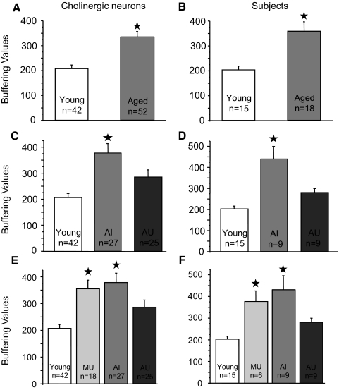
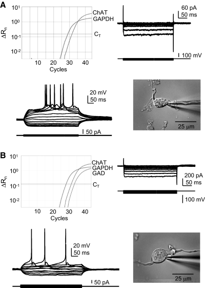
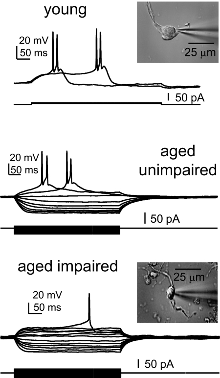
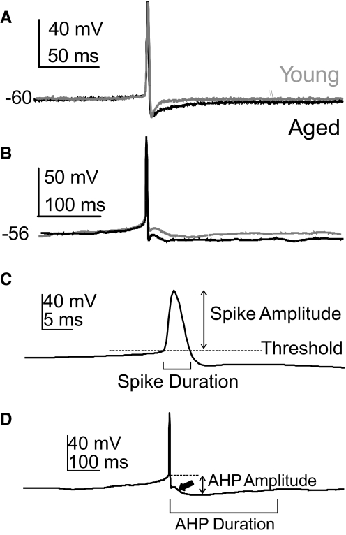
Alterations in neuronal Ca(2+) homeostasis are important determinants of age-related cognitive impairment. We examined the Ca(2+) influx, buffering, and electrophysiology of basal forebrain neurons in adult, middle-aged, and aged male F344 behaviorally assessed rats. Middle-aged and aged rats were characterized as cognitively impaired or unimpaired by water maze performance relative to young cohorts. Patch-clamp experiments were conducted on neurons acutely dissociated from medial septum/nucleus of the diagonal band with post hoc identification of phenotypic marker mRNA using single-cell RT-PCR. We measured whole cell calcium and barium currents and dissected these currents using pharmacological agents. We combined Ca(2+) current recording with Ca(2+)-sensitive ratiometric microfluorimetry to measure Ca(2+) buffering. Additionally, we sought changes in neuronal firing properties using current-clamp recording. There were no age- or cognition-related changes in the amplitudes or fractional compositions of the whole cell Ca(2+) channel currents. However, Ca(2+) buffering was significantly enhanced in cholinergic neurons from aged cognitively impaired rats. Moreover, increased Ca(2+) buffering was present in middle-aged rats that were not cognitively impaired. Firing properties were largely unchanged with age or cognitive status, except for an increase in the slow afterhyperpolarization in aged cholinergic neurons, independent of cognitive status. Furthermore, acutely dissociated basal forebrain neurons in which choline acetyltransferase mRNA was detected had the electrophysiological profiles of identified cholinergic neurons. We conclude that enhanced Ca(2+) buffering by cholinergic basal forebrain neurons may be important during aging.


Figures

Fig. 1.
Fig. 1.
Behavioral testing and assessment of cognitive status of individual F344 rats. Rats were trained in the hidden platform version of the Morris water maze and tested by probe trials as described in methods. A: learning curves of all rats. The graph shows mean cumulative pathlength during each of the successive training trials for young, middle-aged, and aged rats. B: scatterplot shows the Learning Index (LI) score distribution of individual subjects within the 3 age groups for all of the rats in the sample used for electrophysiology experiments. Horizontal lines indicate the mean for each age group. LI scores were calculated as described in methods. C: scatterplot of LIs of rats in the calcium buffering experiments. The horizontal line at the LI score of 280 indicates the cutoff between rats considered cognitively impaired and those considered unimpaired and represents a score that is outside the range of the young animals. Subjects earning an LI score >280 were considered cognitively impaired. The learning curves and mean LI scores of each age group were significantly different from one another, as described in the text.
Fig. 2.
Fig. 2.
Perforated-patch recording of high-voltage–activated (HVA) Ba2+ current densities in cholinergic basal forebrain (BF) neurons from behaviorally characterized rats. Acutely dissociated voltage-clamped BF neurons were assessed for HVA voltage-gated Ca2+ channel (VGCC) function by measuring the peak current density (pA/pF) generated by a standard ramp voltage in perforated-patch configuration with 2 mM Ba2+ as charge carrier. Solutions for the isolation of HVA Ba2+ current are described in methods and neurons were determined to be cholinergic by detection of choline acetyltransferase (ChAT) sequence by post hoc single-cell reverse transcription/polymerase chain reaction (scRT-PCR). A: example HVA current in response to the standard voltage ramp from a holding potential of −80 mV. The arrow indicates the peak inward Ba2+ current. B: graphs of mean Ba2+ current densities by age show that there were no significant age-related differences. C: there were also no significant differences in the HVA current densities between BF cholinergic neurons of subjects with different cognitive status. “n” values refer to the number of cholinergic neurons in the sample. Error bars represent SE. In this and other figures: MU, middle-aged cognitively unimpaired; MI, middle-aged impaired; AU, aged unimpaired; AI, aged impaired.
Fig. 3.
Fig. 3.
Perforated-patch recording of components of the HVA current in cholinergic BF neurons from behaviorally characterized rats. Ba2+ currents were generated in acutely dissociated voltage-clamped neurons by a voltage step from −70 mV to the peak current voltage determined by voltage ramp (as in Fig. 2). Control currents were compared with those in the presence of the HVA antagonists 10 μM nifedipine (blocks L-type current) and 500 nM ω-conotoxin (CTX) MVIIC (blocks N- and P/Q-type currents). A: example of calcium current block by CTX in a BF cholinergic neuron with peak HVA current occurring at −15 mV. The control HVA current is superimposed over the current in ω-CTX MVIIC. B: summary data show that there were no age- or cognition-related differences in the amount of HVA current block by ω-CTX MVIIC. C: summary data for inhibition by nifedipine. There were no significant differences in the amount of block by nifedipine. “n” values refer to the number of cholinergic neurons in the sample. Error bars represent SE.
Fig. 4.
Fig. 4.
Determination of neuronal calcium buffering values in individual cholinergic basal forebrain neurons from behaviorally characterized rats. Acutely dissociated BF neurons were whole cell voltage-clamped at −60 mV and depolarized to 0 mV for several different durations to provide several levels of Ca2+ influx via VGCCs using 2 mM Ca2+ as charge carrier. Simultaneously, the intracellular Ca2+ concentration ([Ca2+]i) in the soma was monitored with fura-2–based ratiometric microfluorimetry. Buffering curves were constructed to relate the Ca2+ influx to the calibrated fura signal as described in methods. Examples of data from single cholinergic neurons are shown for each of the age/cognitive status groups: young (top left), aged unimpaired (top middle), aged impaired (top right), and middle-aged unimpaired (bottom). The data for each cell are composed of a set of superimposed Ca2+ current recordings (top), the corresponding fura-2 fluorescence ratio records with calibrated [Ca2+] (middle), and the buffering curves constructed from the data (bottom). For each neuron, the calculated buffering value (β) is shown, as is the LI score in parentheses for that individual rat. All neurons included in the buffering analysis were found to express ChAT by post hoc scRT-PCR.
Fig. 5.
Fig. 5.
Ca2+ buffering values of cholinergic BF neurons in behaviorally characterized F344 rats during aging. Summary data showing the mean buffering values of all young and aged ChAT+ neurons (A) and the mean buffering values of ChAT+ neurons averaged for each young and aged subject (B). The mean buffering values of cholinergic neurons with the aged subjects divided into impaired and unimpaired cognitive status groups are shown in C, whereas the buffering values averaged within each subject are shown in D. Summary graphs incorporating the mean buffering values of ChAT+ neurons (E) and subjects (F) from middle-aged unimpaired rats are also shown. Note that β values for middle-aged unimpaired neurons and subjects are similar to those of the aged-impaired group. Stars indicate a significant difference (P < 0.05) from starless means. Error bars represent SE and statistical comparisons were by ANOVA for multiple comparisons.
Fig. 6.
Fig. 6.
Properties of ChAT+/GAD+ neurons resemble those of ChAT+ neurons rather than GAD+ neurons. BF neurons in which ChAT sequence is detected by RT-PCR resemble one another and stereotypical BF cholinergic neurons regardless of the detection of GAD sequence. Each example shows the real-time PCR amplification plot (top left), voltage-clamp records of the inward rectifier current in response to hyperpolarizing voltage steps from a holding potential of −60 mV (top right), current-clamp data of superimposed membrane voltage records during a series of small positive and negative current steps from −60 mV (bottom left), and a photo of the acutely dissociated neuron (bottom right). A: ChAT+ neuron. B: ChAT+/GAD+ neuron. Both examples are from aged rats. Current-clamp solutions (see methods) were used for this set of experiments. ChAT, choline acetyltransferase; GAPDH, glyceraldehyde-3-phosphate dehydrogenase; GAD, glutamic acid decarboxylase; CT, detection threshold; ΔRn, change in reporter fluorescence.
Fig. 7.
Fig. 7.
Cholinergic BF neurons display stereotypical properties that remain very similar during aging and are unaffected by cognitive status. Examples shown are from current-clamped ChAT+ BF neurons from a young, an aged cognitively unimpaired and an aged cognitively impaired rat. Each neuron's resting membrane potential (Em) was near −60 mV, and small depolarizing and hyperpolarizing current steps were applied to produce action potentials and inward rectification. Insets are photos of the acutely dissociated neurons.
Fig. 8.
Fig. 8.
The slow afterhyperpolarization (sAHP) duration is increased in aged cholinergic BF neurons. A: superimposed current-clamp records showing an action potential (AP) from a young and an aged cholinergic BF neuron without afterdepolarizations (ADPs: young:gray; aged:black). The spontaneous resting potential is given. B: superimposed current-clamp records as above, except from neurons displaying an ADP. In each case the sAHP of the aged neuron is of longer duration. APs occurred spontaneously without depolarizing current pulses and examples were selected for APs of nearly identical amplitude and duration. Quantitative data are presented in Table 1. C: measurement of the AP amplitude, duration, and threshold is illustrated. Amplitude was measured relative to threshold and duration was measured at threshold. D: an example of the measurement of the sAHP amplitude and duration is shown. Amplitude was measured relative to threshold and duration was measured from the time that the repolarizing phase of the AP crossed the threshold voltage to the time that the potential returned to the pre-AP level. The thick arrow indicates the ADP.

Similar articles

Cited by

References

    1. Allen TG, Abogadie FC, Brown DA. Simultaneous release of glutamate and acetylcholine from single magnocellular “cholinergic” basal forebrain neurons. J Neurosci 26: 1588–1595, 2006 - PMC - PubMed
    1. Bartus RT. On neurodegenerative diseases, models, and treatment strategies: lessons learned and lessons forgotten a generation following the cholinergic hypothesis. Exp Neurol 163: 495–529, 2000 - PubMed
    1. Baxter MG, Frick KM, Price DL, Breckler SJ, Markowska AL, Gorman LK. Presynaptic markers of cholinergic function in the rat brain: relationship with age and cognitive status. Neuroscience 89: 771–780, 1999 - PubMed
    1. Berridge MJ, Lipp P, Bootman MD. The versatility and universality of calcium signalling. Nat Rev Mol Cell Biol 1: 11–21, 2000 - PubMed
    1. Bezprozvanny I, Mattson M. Neuronal calcium mishandling and the pathogenesis of Alzheimer's disease. Trends Neurosci 31: 454–463, 2008 - PMC - PubMed

Publication types

MeSH terms