. 2009 Nov;297(5):C1103-12.
doi: 10.1152/ajpcell.00283.2009. Epub 2009 Aug 12.

Orai1, a critical component of store-operated Ca2+ entry, is functionally associated with Na+/Ca2+ exchanger and plasma membrane Ca2+ pump in proliferating human arterial myocytes

Affiliations

Orai1, a critical component of store-operated Ca2+ entry, is functionally associated with Na+/Ca2+ exchanger and plasma membrane Ca2+ pump in proliferating human arterial myocytes

Sergey G Baryshnikov et al. Am J Physiol Cell Physiol. 2009 Nov.

Abstract

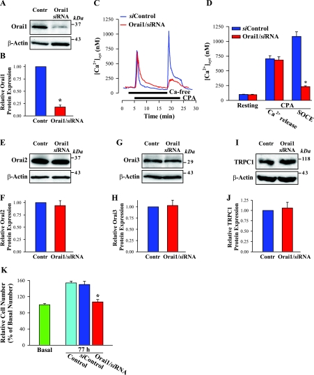
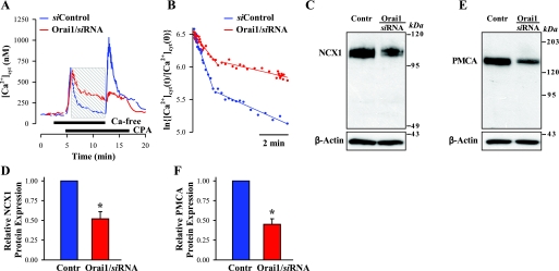
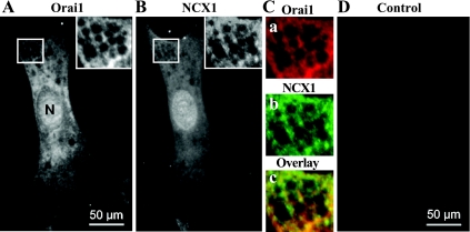
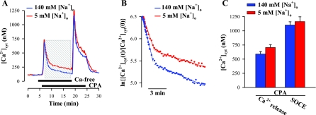
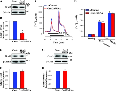
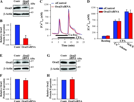
Ca(2+) entry through store-operated channels (SOCs) in the plasma membrane plays an important role in regulation of vascular smooth muscle contraction, tone, and cell proliferation. The C-type transient receptor potential (TRPC) channels have been proposed as major candidates for SOCs in vascular smooth muscle. Recently, two families of transmembrane proteins, Orai [also known as Ca(2+) release-activated Ca(2+) channel modulator (CRACM)] and stromal interacting molecule 1 (STIM1), were shown to be essential for the activation of SOCs mainly in nonexcitable cells. Here, using small interfering RNA, we show that Orai1 plays an essential role in activating store-operated Ca(2+) entry (SOCE) in primary cultured proliferating human aortic smooth muscle cells (hASMCs), whereas Orai2 and Orai3 do not contribute to SOCE. Knockdown of Orai1 protein expression significantly attenuated SOCE. Moreover, inhibition of Orai1 downregulated expression of Na(+)/Ca(2+) exchanger type 1 (NCX1) and plasma membrane Ca(2+) pump isoform 1 (PMCA1). The rate of cytosolic free Ca(2+) concentration decay after Ca(2+) transients in Ca(2+)-free medium was also greatly decreased under these conditions. This reduction of Ca(2+) extrusion, presumably via NCX1 and PMCA1, may be a compensation for the reduced SOCE. Immunocytochemical observations indicate that Orai1 and NCX1 are clustered in plasma membrane microdomains. Cell proliferation was attenuated in hASMCs with disrupted Orai1 expression and reduced SOCE. Thus Orai1 appears to be a critical component of SOCE in proliferating vascular smooth muscle cells, and may therefore be a key player during vascular growth and remodeling.


Figures

Fig. 1.
Fig. 1.
Knockdown of the Orai1 gene markedly reduces store-operated Ca2+ entry (SOCE) and human aortic smooth muscle cell (hASMC) proliferation. A: Western blot showing knockdown of endogenous Orai1 protein in hASMCs treated with Orai1 small interfering (si)RNA. Contr, cells treated with nontargeting siRNA. Membrane proteins (50 μg/lane) were loaded and probed with specific anti-Orai1 antibodies. Blots were later incubated with anti-β-actin antibodies to verify uniform protein loading. B: data are normalized to the amount of β-actin and expressed as means ± SE from 7 Western blots. *P < 0.001 vs. Orai1 protein expression in control cells. C: representative records showing the time course of cytosolic free Ca2+ concentration ([Ca2+]cyt) changes in control hASMC (siControl) and cell treated with Orai1/siRNA. Cyclopiazonic acid (CPA; 10 μM) was applied to the cells in the absence and presence of extracellular Ca2+, as indicated. Nifedipine (10 μM) was applied 10 min before the traces shown and was maintained throughout the experiment. D: summarized data showing resting [Ca2+]cyt, the CPA-induced transient Ca2+ peak in the absence of extracellular Ca2+, and the amplitude of SOCE in control hASMCs and cells treated with Orai1/siRNA. Data are means ± SE (n = 122 cells transfected with nontargeting siRNA and n = 94 cells transfected with Orai1/siRNA, 56 coverslips). *P < 0.001 vs. control. E, G, and I: Western blot analysis of Orai2 (E; 50 μg/lane), Orai3 (G; 30 μg/lane), and C-type transient receptor potential (TRPC)1 (I; 40 μg/lane) protein expression in control hASMCs and cells treated with Orai1/siRNA. F, H, and J: data are normalized to the amount of β-actin and expressed as means ± SE from 4 (F), 6 (H), and 4 (J) Western blots. K: transfection with Orai1/siRNA inhibits hASMC proliferation. Cell numbers were determined before (Basal) and after incubation for 77 h in control growth medium (Control) or medium containing siControl or Orai1/siRNA. Data are presented as % of control (Basal) cell number (100%) and expressed as means ± SE from 4 experiments/transfections. *P < 0.05 vs. siControl.
Fig. 2.
Fig. 2.
Knockdown of the Orai1 gene greatly slows down [Ca2+]cyt decay following CPA-induced Ca2+ transients in Ca2+-free medium and downregulates expression of Na+/Ca2+ exchanger 1(NCX1) and plasma membrane Ca2+ pump 1 (PMCA1) in hASMCs. A: representative records showing the time course of [Ca2+]cyt changes in control hASMC (siControl) and cell treated with Orai1/siRNA. The [Ca2+]cyt decline in the gray boxed portion was fitted to 2 exponentials (Eqs. 1 and 2), and the result of the fitting is plotted in B. B: plot of the fast (τf) and slow (τs) components of [Ca2+]cyt decay for cells treated with siControl and Orai1/siRNA. C and E: Western blot analysis of NCX1 (C; 50 μg/lane) and PMCA1 (E; 30 μg/lane) protein expression in control hASMCs and cells treated with Orai1/siRNA. D and F: data are normalized to the amount of β-actin and expressed as means ± SE from 5 (D) and 5 (F) Western blots. *P < 0.001 vs. control.
Fig. 3.
Fig. 3.
Immunofluorescent localization of Orai1 and NCX1 in hASMC. A and B: images of cell double labeled with anti-Orai1 antibody (A) and anti-NCX1 antibody (B). Insets in A and B (enlargements of boxed areas) indicate that Orai1 and NCX1 labels show similar distributions. C, a and b: pseudocolor images (red, anti-Orai1; green, anti-NCX1) of enlarged boxes from A and B, respectively. c: Colocalization of Orai1 (a) and NCX1 (b) staining; yellow, orange, and yellow/green areas in overlay indicate regions of overlap between the 2 epitopes. D: fluorescence detected from the secondary antibody (Cy3) in the absence of primary anti-Orai1 antibody (control). Scale bars, 50 μm (A, D). N, nucleus. Similar results were obtained in 17 cells.
Fig. 4.
Fig. 4.
NCX does not contribute to SOCE in hASMCs. A: representative records showing time course of [Ca2+]cyt changes in response to CPA (10 μM) in absence and presence of extracellular Ca2+. hASMCs were superfused with solutions containing 140 mM or 5 mM extracellular Na+ concentration ([Na+]o). The [Ca2+]cyt decline in the gray boxed portion was fitted to 2 exponentials (Eqs. 1 and 2), and the result of the fitting is plotted in B. Nifedipine (10 μM) was applied 10 min before the traces shown and was maintained throughout the experiment. B: plot of τf and τs of [Ca2+]cyt decay at 140 mM [Na+]o and 5 mM [Na+]o. Values of τf and τs were significantly larger in cells perfused with solution containing 5 mM Na+ (conditions that block NCX1-mediated Ca2+ extrusion) than in cells bathed in control solution (140 mM Na+) (2.55 and 23.81 min vs. 1.78 and 17.54 min, respectively). C: summarized data showing the amplitude of SOCE in cells bathed in 140 mM Na+ solution or in 5 mM Na+ solution (n = 44 and 48 cells, respectively; 10 coverslips).
Fig. 5.
Fig. 5.
Knockdown of the Orai2 gene does not affect SOCE in hASMCs. A: Western blot of Orai2 expression in hASMCs treated with siControl (Contr) and Orai2/siRNA. Membrane proteins (10 μg/lane) were loaded and probed with specific anti-Orai2 antibodies. Blots were later incubated with anti-β-actin antibodies to verify uniform protein loading. B: data are normalized to the amount of β-actin and expressed as means ± SE from 4 Western blots. *P < 0.001 vs. Orai2 protein expression in control cells. C: representative records showing the time course of [Ca2+]cyt changes in control hASMC (siControl) and cell treated with Orai2/siRNA. CPA (10 μM) was applied to the cells in the absence and presence of extracellular Ca2+, as indicated. Nifedipine (10 μM) was applied 10 min before the traces shown and was maintained throughout the experiment. D: summarized data showing resting [Ca2+]cyt, the CPA-induced transient Ca2+ peak in the absence of extracellular Ca2+, and the amplitude of SOCE in control hASMCs (n = 88 cells) and cells treated with Orai2/siRNA (81 cells). Data are means ± SE (n = 24 coverslips). E and G: Western blot analysis of Orai1 (E; 20 μg/lane) and Orai3 (G; 50 μg/lane) protein expression in control hASMCs and cells treated with Orai2/siRNA. F and H: data are normalized to the amount of β-actin and expressed as means ± SE from 4 (F) and 4 (H) Western blots.
Fig. 6.
Fig. 6.
Effect of knockdown of Orai3 on SOCE in hASMCs. A: Western blot showing knockdown of endogenous Orai3 protein in cells treated with Orai3/siRNA. Contr, cells treated with nontargeting siRNA. Membrane proteins (30 μg/lane) were loaded and probed with specific anti-Orai3 antibodies. B: data are normalized to the amount of β-actin and expressed as means ± SE from 10 Western blots. *P < 0.001 vs. Orai3 protein expression in control cells. C: representative records showing time course of [Ca2+]cyt changes in control hASMC (siControl) and cell treated with Orai3/siRNA. CPA (10 μM) was applied to the cells in the absence and presence of extracellular Ca2+, as indicated. Nifedipine (10 μM) was applied 10 min before the traces shown and was maintained throughout the experiment. D: summarized data showing resting [Ca2+]cyt, the CPA-induced transient Ca2+ peak in the absence of extracellular Ca2+, and the amplitude of SOCE in control hASMCs and cells treated with Orai3/siRNA. Data are means ± SE (n = 94 cells; 28 coverslips). E and G: Western blot analysis of Orai1 (E; 30 μg/lane) and Orai2 (G; 20 μg/lane) protein expression in hASMCs treated with siControl and Orai3/siRNA. F and H: Data are normalized to the amount of β-actin and expressed as means ± SE from 7 (F) and 4 (H) Western blots.

Similar articles

Cited by

References

    1. Akaike N, Kostyuk PG, Osipchuk YV. Dihydropyridine-sensitive low-threshold calcium channels in isolated rat hypothalamic neurones. J Physiol 412: 181–195, 1989 - PMC - PubMed
    1. Albert AP, Large WA. Store-operated Ca2+-permeable non-selective cation channels in smooth muscle cells. Cell Calcium 33: 345–356, 2003 - PubMed
    1. Albert AP, Saleh SN, Peppiatt-Wildman CM, Large WA. Multiple activation mechanisms of store-operated TRPC channels in smooth muscle cells. J Physiol 583: 25–36, 2007 - PMC - PubMed
    1. Ambudkar IS, Ong HL, Liu X, Bandyopadhyay BC, Cheng KT. TRPC1: the link between functionally distinct store-operated calcium channels. Cell Calcium 42: 213–223, 2007 - PubMed
    1. Beech DJ. Emerging functions of 10 types of TRP cationic channel in vascular smooth muscle. Clin Exp Pharmacol Physiol 32: 597–603, 2005 - PMC - PubMed

Publication types

MeSH terms

LinkOut - more resources