

Case Reports
doi: 10.1056/NEJMc0903652.
Vision 1 year after gene therapy for Leber's congenital amaurosis
- PMID: 19675341
- PMCID: PMC2847775
- DOI: 10.1056/NEJMc0903652
Item in Clipboard
Case Reports
Vision 1 year after gene therapy for Leber's congenital amaurosis
N Engl J Med.
.
No abstract available
Figures
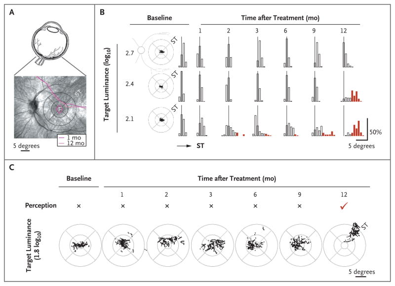
Panel A shows the eye (upper image) and the patient’s retina (lower image). Overlaid contours of constant sensitivity (measured by means of microperimetry) show no change in visual sensitivity between 1 and 12 months after treatment. F denotes the fovea, and ST the superotemporal retina that received treatment. The circular pattern is a standard grid centered on the fovea. Retinal distance calibration corresponding to 5 degrees of visual angle is shown. Panel B shows fixation clouds (scatter plots) in the study eye of the patient at baseline and the statistics of fixation dwell time (bar graphs) along the diagonal meridian as a function of the target luminance. All three luminances were perceived by the patient at all visits. At the 2.7- and 2.4-log10 luminances, fixation was within 3 degrees of the fovea more than 99% of the time at all visits except at the 12-month visit for 2.4-log10 luminance, when 68% of fixation time dwelled in an ST retinal region 4 to 9 degrees from the fovea. At 2.1-log10 luminance, fixations showed increasingly greater excursions into the ST retina between 2 and 9 months after treatment. At 12 months, 89% of fixation time dwelled in the ST region 4 to 9 degrees from the fovea. Thin vertical lines represent the foveal location. Red bars indicate significant (>3 degrees) excursions from the fovea. Panel C shows that a dimmer target (1.8 log10) was not perceived by the patient’s study eye during baseline though 9 months after treatment. At 12 months, this stimulus was perceived for the first time with a coincident shift of fixation into the ST retinal region.
References
-
- den Hollander AI, Roepman R, Koenekoop RK, Cremers FP. Leber congenital amaurosis: genes, proteins and disease mechanisms. Prog Retin Eye Res. 2008;27:391–419. - PubMed
-
- Bainbridge JWB, Smith AJ, Barker SS, et al. Effect of gene therapy on visual function in Leber’s congenital amaurosis. N Engl J Med. 2008;358:2231–9. - PubMed
Publication types
MeSH terms
Grants and funding
LinkOut - more resources
Full Text Sources
Other Literature Sources
Medical