Review
. 2010 Jan;35(1):27-47.
doi: 10.1038/npp.2009.93.

Cortico-Basal Ganglia reward network: microcircuitry

Affiliations
Review

Cortico-Basal Ganglia reward network: microcircuitry

Susan R Sesack et al. Neuropsychopharmacology. 2010 Jan.

Abstract

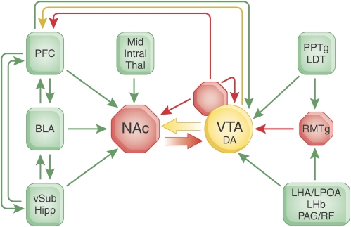
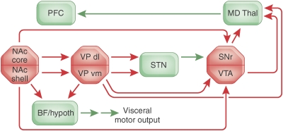
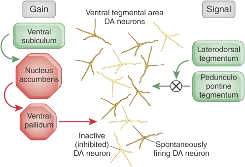
Many of the brain's reward systems converge on the nucleus accumbens, a region richly innervated by excitatory, inhibitory, and modulatory afferents representing the circuitry necessary for selecting adaptive motivated behaviors. The ventral subiculum of the hippocampus provides contextual and spatial information, the basolateral amygdala conveys affective influence, and the prefrontal cortex provides an integrative impact on goal-directed behavior. The balance of these afferents is under the modulatory influence of dopamine neurons in the ventral tegmental area. This midbrain region receives its own complex mix of excitatory and inhibitory inputs, some of which have only recently been identified. Such afferent regulation positions the dopamine system to bias goal-directed behavior based on internal drives and environmental contingencies. Conditions that result in reward promote phasic dopamine release, which serves to maintain ongoing behavior by selectively potentiating ventral subicular drive to the accumbens. Behaviors that fail to produce an expected reward decrease dopamine transmission, which favors prefrontal cortical-driven switching to new behavioral strategies. As such, the limbic reward system is designed to optimize action plans for maximizing reward outcomes. This system can be commandeered by drugs of abuse or psychiatric disorders, resulting in inappropriate behaviors that sustain failed reward strategies. A fuller appreciation of the circuitry interconnecting the nucleus accumbens and ventral tegmental area should serve to advance discovery of new treatment options for these conditions.


Figures

Figure 1
Figure 1
Principal afferents linking brain centers for goal-directed behavior with the NAc and VTA. For clarity, only some of the projections are shown, and the principal efferent pathways from the NAc are illustrated in Figure 2. Red indicates inhibitory structures and pathways, green excitatory connections, and yellow the modulatory influence of DA. Please refer to the text for detailed explanation. BLA, basolateral amygdala; LHA/LPOA, lateral hypothalamic and lateral preoptic areas; LHb, lateral habenula; Mid/Intral Thal, midline and intralaminar thalamic nuclei; NAc, nucleus accumbens; PAG/RF, periaqueductal gray and reticular formation; PFC, prefrontal cortex; PPTg/LDT, pedunculopontine and laterodorsal tegmentum; RMTg, mesopontine rostromedial tegmental nucleus; VP, ventral pallidum; vSub/Hipp, ventral subiculum of the hippocampus; VTA, ventral tegmental area.
Figure 2
Figure 2
Hypothetical direct and indirect output pathways whereby the NAc core and shell may disinhibit or inhibit, respectively, adaptive motor pathways for maximizing reward acquisition. Only major projections are shown. Red indicates inhibitory structures and pathways, whereas green indicates excitatory connections. Please refer ‘Efferents' in section Nucleus Accumbens for detailed explanation. BF Hypoth, basal forebrain and hypothalamus; MD Thal, mediodorsal thalamic nucleus; NAc, nucleus accumbens; PFC, prefrontal cortex; SNr, substantia nigra zona reticulata; STN, subthalamic nucleus; VP dl/vm, ventral pallidum, dorsolateral, and ventromedial; VTA, ventral tegmental area.
Figure 3
Figure 3
DA neurons in the VTA can exist in several activity states. In the basal, unstimulated state, DA neurons fire spontaneously at a slow, irregular rate. The VP provides a potent GABAergic input to DA neurons, causing a proportion of them to be tonically inhibited and non-firing. The VP in turn is controlled by afferents from the vSub and the NAc. When the vSub is activated, it provides a glutamatergic drive to the NAc, which in turn inhibits the VP and releases DA neurons from inhibition, allowing them to fire spontaneously. In contrast, the PPTg provides a potent direct glutamatergic input to DA neurons; when the PPTg is activated, it causes DA neurons to fire in bursts, which is believed to be the behaviorally salient pattern signaling a rewarding event. The impact of the PPTg, however, is gated by the LDT; only when the LDT is active can the PPTg initiate burst firing. In order for a DA neuron to burst fire, it must first be firing spontaneously. Given that the vSub controls the proportion of DA neurons firing spontaneously, it also sets the number of DA neurons that can be made to burst fire by the PPTg. As such, the PPTg drives the behaviorally salient burst firing, whereas the vSub provides the ‘gain' or amplification of the signal. The greater the vSub-driven gain, the larger the DA response produced by a stimulus that activates the PPTg.

Similar articles

Cited by

References

    1. Alcantara AA, Chen V, Herring BE, Mendenhall JM, Berlanga ML. Localization of dopamine D2 receptors on cholinergic interneurons of the dorsal striatum and nucleus accumbens of the rat. Brain Res. 2003;986:22–29. - PubMed
    1. Alexander GE, Crutcher MD, DeLong MR. Basal ganglia–thalamocortical circuits: parallel substrates for motor, oculomotor, ‘prefrontal' and ‘limbic' functions. Prog Brain Res. 1990;85:119–146. - PubMed
    1. Alheid GF, Heimer L. New perspectives in basal forebrain organization of special relevance for neuropsychiatric disorders: the striatopallidal, amygdaloid, and corticopetal components of substantia innominata. Neuroscience. 1988;27:1–39. - PubMed
    1. Amaral DG, Dolorfo C, Alvarez-Royo P. Organization of CA1 projections to the subiculum: a PHA-L analysis in the rat. Hippocampus. 1991;1:415–435. - PubMed
    1. Ambroggi F, Ishikawa A, Fields HL, Nicola SM. Basolateral amygdala neurons facilitate reward-seeking behavior by exciting nucleus accumbens neurons. Neuron. 2008;59:648–661. - PMC - PubMed

Publication types

MeSH terms