. 2009 Sep 17;461(7262):361-6.
doi: 10.1038/nature08319. Epub 2009 Aug 12.

The structural basis of tail-anchored membrane protein recognition by Get3

Affiliations

The structural basis of tail-anchored membrane protein recognition by Get3

Agnieszka Mateja et al. Nature. .

Abstract

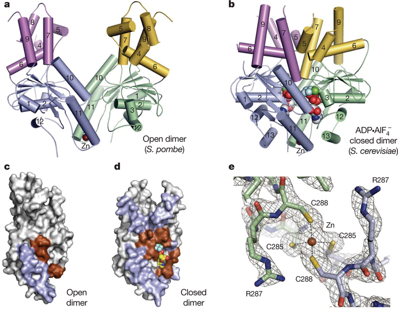
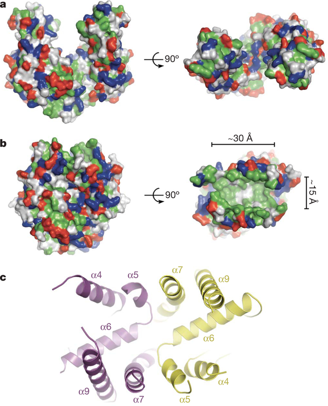
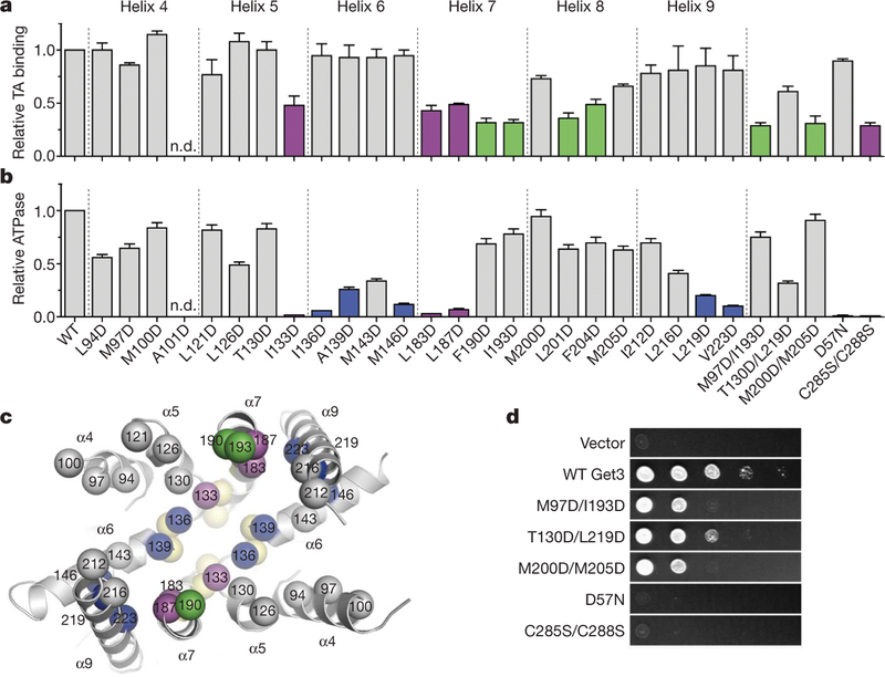
Targeting of newly synthesized membrane proteins to the endoplasmic reticulum is an essential cellular process. Most membrane proteins are recognized and targeted co-translationally by the signal recognition particle. However, nearly 5% of membrane proteins are 'tail-anchored' by a single carboxy-terminal transmembrane domain that cannot access the co-translational pathway. Instead, tail-anchored proteins are targeted post-translationally by a conserved ATPase termed Get3. The mechanistic basis for tail-anchored protein recognition or targeting by Get3 is not known. Here we present crystal structures of yeast Get3 in 'open' (nucleotide-free) and 'closed' (ADP.AlF(4)(-)-bound) dimer states. In the closed state, the dimer interface of Get3 contains an enormous hydrophobic groove implicated by mutational analyses in tail-anchored protein binding. In the open state, Get3 undergoes a striking rearrangement that disrupts the groove and shields its hydrophobic surfaces. These data provide a molecular mechanism for nucleotide-regulated binding and release of tail-anchored proteins during their membrane targeting by Get3.


Figures

Figure 1 |
Figure 1 |. Get3 is a dynamic, metal-stabilized homodimer.
a, b, Crystal structures of Get3 in open (a) and closed (b) dimer states. Each monomer comprises a core ATPase subdomain (blue, green) and an α-helical subdomain (magenta, yellow). A tightly bound zinc atom (brown sphere) lies at the dimer interface. c, d, Residues in the homodimer interface (blue, brown) are mapped to the surface of a Get3 monomer from the open (c) and closed (d) dimer structures. Monomers are rotated ~90° about the dimer pseudo-two-fold axis, relative to a and b. Interfacial regions involving the conserved ATPase motifs are coloured brown. e, Details of the Get3 dimerization motif in the closed dimer structure, highlighting the tetrahedral coordination of zinc by the conserved CXXC sequence motif. Electron density is from a σA-weighted 2FoFc map calculated at 2.0 Å resolution and contoured at 2σ.
Figure 2 |
Figure 2 |. A composite hydrophobic groove at the closed dimer interface.
a, Surface representation of the Get3 open dimer with hydrophobic residues coloured green; positively and negatively charged residues are coloured blue and red, respectively. b, As in a, but for the Get3 closed dimer. The approximate dimensions of the large hydrophobic groove (right panel) are indicated. c, Architecture of the composite hydrophobic groove formed by the association of α-helical subdomains (magenta and yellow, coloured as in Fig. 1 and oriented as in b, right panel) at the homodimer interface.
Figure 3 |
Figure 3 |. Functional analysis of the hydrophobic groove.
a, The effect of site-specific Get3 mutations on binding to full-length human SEC61β was measured by native immunoprecipitation. Each value is the average of between three and six independent measurements, performed on different days, relative to wild type. Error bars denote s.e.m. Variants showing less than ~50% of wild-type binding are highlighted (green, magenta). b, ATPase activity was determined in triplicate, and values are Vmax relative to wild type. Error bars denote s.e.m. Variants showing less than ~25% of wild-type ATPase activity are highlighted (blue, magenta). c, Mutations showing the strongest defects in TA substrate binding (green), ATP hydrolysis (blue), or both (magenta) are mapped onto the composite hydrophobic groove (oriented as in Fig. 2c). ATPase mutants localize to the base of the groove, adjacent to the Get3 nucleotide sensor (yellow; see Fig. 4b). Helix α8 is disordered in the Get3 closed dimer structure and is therefore not visible here. d, In vivo analysis of Get3 mutants. WT, wild type.
Figure 4 |
Figure 4 |. The Get3 nucleotide sensor.
a, Key interactions within the composite ATP-binding site of one subunit (green) of the closed dimer. The essential catalytic residue Asp 57 coordinates the putative nucleophilic water molecule (red sphere, asterisk), adjacent to AlF4. The nucleotide makes further interactions with residues in the second subunit (blue), including the P-loop residue Lys 26. b, A coil-to-helix transition in the Switch II region (green and blue) is observed in the presence of ADP·AlF 4 relative to the nucleotide free state (grey subunit). Viewed ~180° from the orientation in c, looking along the dimer pseudo-two-fold axis, and coloured as in Fig. 1. Conserved, cross-monomer interactions between Switch II/α7 and the α-helical subdomain are disrupted in the open dimer (the second subunit of the nucleotide-free dimer is not visible here). c, The α-helical subdomains move apart in the open dimer (grey), and helices within each of the resulting ‘half-sites’ rearrange; helix α8, disordered in the closed dimer, inserts into the hydrophobic half-site to shield it from solvent.
Figure 5 |
Figure 5 |. Model for TA protein targeting.
a, The 20-residue α-helical TMD of the Methanococcus jannaschii TA protein Sec61β (PDB accession 1RHZ) can be modelled into the Get3 hydrophobic groove with good physiochemical complementarity. b, The Get3 open dimer binds ATP and newly synthesized TA proteins destined for the ER (step 1). The Get3-substrate complex is targeted to the ER by an interaction with the membrane bound receptor, Get1/2 (step 2). After ATP hydrolysis, conformational changes in the nucleotide sensor destabilize the composite groove, driving TA substrate release (step 3). Membrane insertion may be spontaneous, or facilitated by a dedicated integrase (not shown). Disruption of the closed dimer following ATP hydrolysis and TA substrate insertion drives release from the membrane and restores Get3 to its open dimer configuration (step 4).

References

    1. Egea PF, Stroud RM & Walter P Targeting proteins to membranes: structure of the signal recognition particle. Curr. Opin. Struct. Biol 15, 213–220 (2005). - PubMed
    1. Keenan RJ, Freymann DM, Stroud RM & Walter P The signal recognition particle. Annu. Rev. Biochem 70, 755–775 (2001). - PubMed
    1. Rapoport TA Protein translocation across the eukaryotic endoplasmic reticulum and bacterial plasma membranes. Nature 450, 663–669 (2007). - PubMed
    1. Beilharz T, Egan B, Silver PA, Hofmann K & Lithgow T Bipartite signals mediate subcellular targeting of tail-anchored membrane proteins in Saccharomyces cerevisiae. J. Biol. Chem 278, 8219–8223 (2003). - PubMed
    1. Kalbfleisch T, Cambon A & Wattenberg BW A bioinformatics approach to identifying tail-anchored proteins in the human genome. Traffic 8, 1687–1694 (2007). - PubMed

Publication types

MeSH terms