

Regeneration of multilineage skin epithelia by differentiated keratinocytes
- PMID: 19675579
- PMCID: PMC2879264
- DOI: 10.1038/jid.2009.244
Regeneration of multilineage skin epithelia by differentiated keratinocytes
Abstract
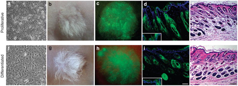
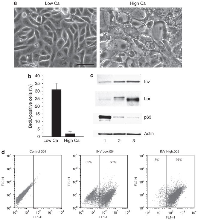
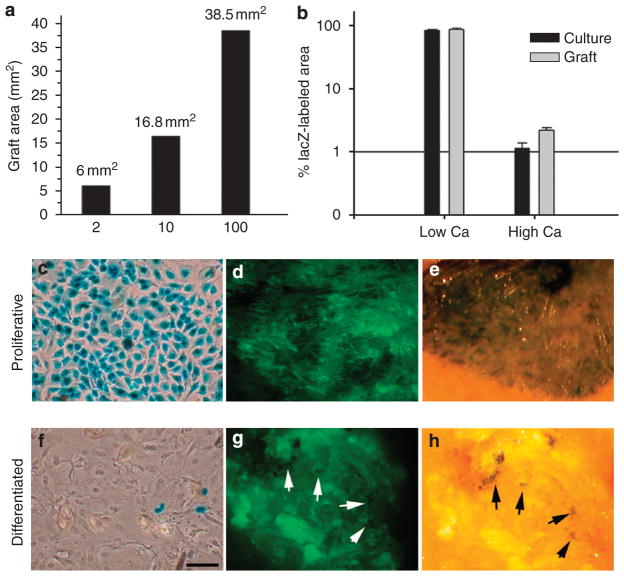
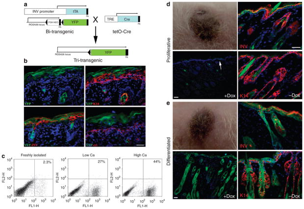
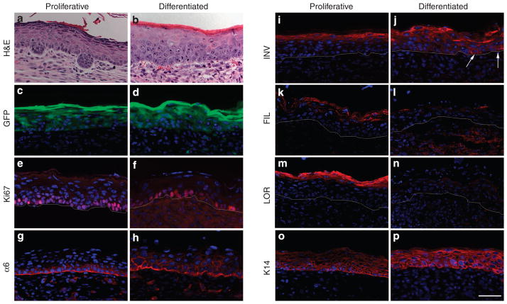
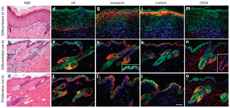
Although homeostasis of rapidly renewing tissues like skin epithelia is maintained by stem cells, the committed progeny of stem cells in the basal layer of epidermis retain regenerative potential and are capable of forming epidermis in response to environmental cues. It is not clear, however, at what point within the epidermal lineage keratinocytes lose this regenerative potential. In this study, we examined the extent of tissue formation by post-mitotic differentiated keratinocytes. We show that cultures of mouse keratinocytes that were, by all measures, differentiated were able to reform a self-renewing, hair-bearing skin when transplanted onto suitable sites in vivo. Genetic labeling and lineage-tracing studies in combination with an involucrin-driven Cre/lox reporter system confirmed that transplanted differentiated keratinocytes were indeed the source of the regenerated skin. More importantly, analysis of early stages of skin regeneration showed hallmarks of dedifferentiation of transplanted differentiated keratinocytes. These data indicate that commitment to differentiation does not prohibit cells from re-entering the cell cycle, de-differentiating, and acquiring "stemness". These findings suggest that epidermis can use different strategies for homeostasis and tissue regeneration.
Conflict of interest statement
The authors state no conflict of interest.
Figures
Similar articles
-
Ectopic expression of the nude gene induces hyperproliferation and defects in differentiation: implications for the self-renewal of cutaneous epithelia.Dev Biol. 1999 Aug 1;212(1):54-67. doi: 10.1006/dbio.1999.9328. Dev Biol. 1999. PMID: 10419685
-
Modulation of the differentiated phenotype of keratinocytes of the hair follicle and from epidermis.J Dermatol Sci. 1994 Jul;7 Suppl:S142-51. doi: 10.1016/0923-1811(94)90045-0. J Dermatol Sci. 1994. PMID: 7999672 Review.
-
Differentiated Daughter Cells Regulate Stem Cell Proliferation and Fate through Intra-tissue Tension.Cell Stem Cell. 2021 Mar 4;28(3):436-452.e5. doi: 10.1016/j.stem.2020.11.002. Epub 2020 Dec 1. Cell Stem Cell. 2021. PMID: 33264636 Free PMC article.
-
Self-assembly of differentiated progenitor cells facilitates spheroid human skin organoid formation and planar skin regeneration.Theranostics. 2021 Jul 25;11(17):8430-8447. doi: 10.7150/thno.59661. eCollection 2021. Theranostics. 2021. PMID: 34373751 Free PMC article.
-
The basement membrane in epidermal polarity, stemness, and regeneration.Am J Physiol Cell Physiol. 2022 Dec 1;323(6):C1807-C1822. doi: 10.1152/ajpcell.00069.2022. Epub 2022 Nov 14. Am J Physiol Cell Physiol. 2022. PMID: 36374168 Review.
Cited by
-
A keratin 15 containing stem cell population from the hair follicle contributes to squamous papilloma development in the mouse.Mol Carcinog. 2013 Oct;52(10):751-9. doi: 10.1002/mc.21896. Epub 2012 Mar 16. Mol Carcinog. 2013. PMID: 22431489 Free PMC article.
-
Defining stem cell dynamics and migration during wound healing in mouse skin epidermis.Nat Commun. 2017 Mar 1;8:14684. doi: 10.1038/ncomms14684. Nat Commun. 2017. PMID: 28248284 Free PMC article.
-
Targeting the stem cell niche: role of collagen XVII in skin aging and wound repair.Theranostics. 2022 Sep 6;12(15):6446-6454. doi: 10.7150/thno.78016. eCollection 2022. Theranostics. 2022. PMID: 36185608 Free PMC article. Review.
-
Protein kinase D is implicated in the reversible commitment to differentiation in primary cultures of mouse keratinocytes.J Biol Chem. 2010 Jul 23;285(30):23387-97. doi: 10.1074/jbc.M110.105619. Epub 2010 May 12. J Biol Chem. 2010. PMID: 20463010 Free PMC article.
-
Human Basal and Suprabasal Keratinocytes Are Both Able to Generate and Maintain Dermo-Epidermal Skin Substitutes in Long-Term In Vivo Experiments.Cells. 2022 Jul 9;11(14):2156. doi: 10.3390/cells11142156. Cells. 2022. PMID: 35883599 Free PMC article.
References
-
- Blanpain C, Lowry WE, Geoghegan A, Polak L, Fuchs E. Self-renewal, multipotency, and the existence of two cell populations within an epithelial stem cell niche. Cell. 2004;118:635–48. - PubMed
Publication types
MeSH terms
Substances
Grants and funding
LinkOut - more resources
Full Text Sources
Molecular Biology Databases
